


Feline viral dermatoses have long remained in the background, partly due to the lack of easily identifiable causative agents and partly due to the absence of specific treatments. Long known for its respiratory and ocular manifestations, feline herpesvirus type 1 is now recognized as a cause of ulcerative dermatitis, primarily affecting the bridge of the nose.
The condition nonetheless remains difficult to diagnose, especially since it can easily mimic allergic dermatitis, and even harder to treat. Let us therefore provide a comprehensive overview of the latest etiopathogenic, diagnostic, and therapeutic knowledge, including the most recent treatments and protocols.
1 The Virus and Its Cycle
1.1 An Epitheliotropic Alphaherpesvirus
Feline herpesvirus type 1 is a double-stranded DNA alphaherpesvirus of which only one serotype exists (Gaskell et al., 2007). It replicates in the nucleus of the cells it infects, producing intranuclear inclusion bodies that mark its presence (Gutzwiller et al., 2007). Its primary target is the epithelium of the upper respiratory tract and the conjunctiva, which is why it is known to all practitioners as the causative agent of feline viral rhinotracheitis. However, its clinical manifestations extend well beyond this framework. Indeed, it can also cause dendritic ulcerative keratitis, eosinophilic keratoconjunctivitis, corneal sequestrum, or anterior uveitis (Persico et al., 2011). The skin can be added to this list.
Recent virological nomenclature designates the agent under the name Felid alphaherpesvirus 1, a denomination adopted by several dermatopathology publications, with the most recent binomial being Varicellovirus felidalpha1, within the family Orthoherpesviridae (Sebastian Pineda et al., 2026a). The abbreviations FHV-1 and FeHV-1, which are older, remain in common use in the literature.
1.2 Ganglionic Latency and Reactivation
The epidemiological behavior of the virus is that of an alphaherpesvirus. Clinically recovered cats become latent carriers and experience periodic reactivation episodes, particularly following stress. The primary site of latency is the trigeminal ganglion (Gaskell et al., 2007). Latency is characterized by low but persistent transcriptional activity and minimal assembly of viral particles, which is sufficient to sustain the host’s immune response and to contain recurrences (Persico et al., 2011). It is estimated that 80% of recovered cats remain latent carriers (Miller et al., 2013). Whenever the immune response weakens, viral replication resumes and lesions may reappear.
Viral shedding accompanies these reactivations. It occurs through oronasal and conjunctival secretions, can last three weeks, and transmission therefore requires direct contact with a shedding cat. Stress and the administration of corticosteroids are the two best-documented circumstances of reactivation (Thiry et al., 2009).
1.3 The Pathway to the Skin
The mechanism by which the virus reaches the skin remains poorly understood. After replication at the inoculation site, feline herpesvirus travels along the facial nerves and establishes latency in the trigeminal ganglion, the optic nerves, the chiasm, the olfactory bulb, and the cornea, but it is not thought to persist in the skin (Gutzwiller et al., 2007). Two hypotheses therefore remain open, neither of which has been proven. The first is ganglionic reactivation followed by centrifugal transport to the cutaneous territories innervated by the trigeminal nerve, which would explain the preferential facial topography. The second is direct inoculation of the epidermis by oronasal secretions, facilitated by scratching and excoriations. The hypothesis currently favored in reference textbooks is that of a recurrence of latent infection within the cutaneous nerves (Munday and Wilhelm, 2020). Stress and cutaneous trauma are moreover presented as factors liable to precipitate the appearance of ulcers, which supports a local component in triggering the condition (Miller et al., 2013). In most published case series, lesions appeared in circumstances suggesting reactivation of latent infection rather than primary infection (Hargis et al., 1999).
One laboratory argument does, however, support replication in the skin itself: viral entry by endocytosis has been observed in primary feline cutaneous fibroblasts, chosen precisely because they constitute a natural target of infection (Synowiec et al., 2023).
The eosinophilic infiltrate, which dominates the histological lesion, remains entirely unexplained. It is unknown why some viral infections recruit eosinophils while others do not; nor is it known whether this recruitment depends directly on the virus or simply reflects keratin released into the dermis by the destruction of adnexa. Three mechanisms have been proposed, none of which has been proven: the production, by infected epithelial cells, of chemotactic cytokines for eosinophils; the perpetuation of recruitment by cytokines produced by the eosinophils themselves; or the synthesis, by the virus, of chemotactic proteins. The authors also note that eosinophilic infiltration is not specific to this infection, as it has been documented in influenza virus, rhinovirus, and respiratory syncytial virus infections (Hargis et al., 1999).
2 Epidemiology and Circumstances of Onset
2.1 A Rare, or Underdiagnosed, Dermatosis
Although the original authors had considered it relatively common, having collected ten cases over only the years 1996 and 1997 (Hargis et al., 1999), studies conducted since then agree on a low prevalence. In one study conducted on thirty cases of eosinophilic granuloma complex, other eosinophilic dermatoses, or stomatitis, only two, or 6.6%, proved positive by immunohistochemistry (Lee et al., 2010). In another study, out of sixty-four clinically compatible biopsies, only two were positive by both PCR and immunohistochemistry (Persico et al., 2011).
2.2 Signalment
No predisposition has been identified. In the most recent case series, there were five castrated males and three spayed females, seven domestic shorthairs and one medium-haired cat, ranging in age from six months to ten years, with a median of eight and a half years (Sebastian Pineda et al., 2026b). Individual published cases cover the same spectrum: a twelve-year-old Bengal and a three-year-old Siamese (Sánchez et al., 2012), a ten-month-old domestic shorthair (Porcellato et al., 2018), and a six-year-old domestic shorthair (Sebastian Pineda et al., 2026a). Age is the only parameter for which a trend emerges: the condition appears more common around five years of age, with cases reported from four months to seventeen years (Munday and Wilhelm, 2020). It is recognized mainly in adults but can also affect kittens (Miller et al., 2013).
2.3 Triggering Circumstances
In the original case series, eight out of ten cats showed no improvement, or even worsened, with systemic corticosteroids, and six of those eight had received recent or concomitant corticosteroid therapy before lesions appeared (Hargis et al., 1999). One case of auricular pinna dermatitis worsened following corticosteroid administration (Porcellato et al., 2018). Lack of response to corticosteroids should therefore be considered a major warning sign in the presence of an ulcerative nasal bridge lesion in a cat (Lee et al., 2010).
Stress associated with overcrowding comes next, particularly in group housing settings, catteries, or shelters. In the original case series, five out of ten cats had a history of chronic or recurrent upper respiratory tract disease, whereas no association was found with retroviral immunosuppression, recent vaccination, or the use of intranasal vaccines (Hargis et al., 1999). It should be kept in mind that prolonged immunomodulatory treatment may play the same role: ciclosporin administered at high doses to immunocompetent cats impairs the primary immune response (Roberts et al., 2015). The specific reactivating effect of ciclosporin was evaluated in cats experimentally infected several months earlier, divided among intramuscular methylprednisolone acetate, oral ciclosporin, and placebo. Signs of reactivation appeared in some animals, but the disease remained mild and spontaneously resolving in most (Lappin and Roycroft, 2015).
3 Clinical Presentation
3.1 The Classic Facial Form
Lesions are located on the nasal planum and on the haired skin of the face, with a predilection for the dorsal and lateral nasal bridge and the periorbital region (Persico et al., 2011). They present as vesicles, erosions, ulcers, and crusts, accompanied by erythema, edema, and exudate of variable intensity; pruritus is mild or even absent (Persico et al., 2011). The appearance is that of an ulcerative and crusted dermatosis, often progressive and persistent, the healing of which may leave a scar (Hargis et al., 1999). In a recent study, the nose was affected in six out of eight cases (Sebastian Pineda et al., 2026b).
Lesions are most often asymmetric, but a symmetric distribution does not rule out a herpetic origin, and regional lymphadenomegaly may accompany them; extension to the entire body within a few days remains exceptional (Munday and Wilhelm, 2020). Ulcers are moreover described as typically superficial and multiple, and they can occur anywhere, including on the paw pads (Miller et al., 2013).
Pruritus varies and it is often what leads toward a diagnosis of allergic dermatitis: mild or even absent (Persico et al., 2011; Albanese, 2017); slight and mainly present at onset (Nuttall et al., 2019); or frankly intense (Munday and Wilhelm, 2020). Pruritus therefore neither supports nor rules out the herpetic hypothesis.
Chronicity is the rule rather than the exception. A nasal lesion persisted for at least seven months under topical treatments, before almost completely regressing without treatment, then recurring at the same site and reaching its original size, for a total duration of fifteen months (Hargis et al., 1999). The disease therefore follows a relapsing course, punctuated by spontaneous remissions.
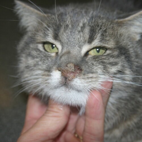
Classic manifestations of feline cutaneous herpesvirus infection
3.2 Extrafacial Forms
These are rare but well documented, and failure to recognize them is a direct source of diagnostic error. Two cats, a twelve-year-old Bengal and a three-year-old Siamese, presented with plaques and ulcers on the flank and inner thigh, with no facial lesions whatsoever (Sánchez et al., 2012). Chronic unilateral ulcerative dermatitis of the left auricular pinna, with adherent crusts, had been evolving for six months in another ten-month-old cat (Porcellato et al., 2018). Similarly, a case was described of a six-year-old cat presenting with lesions on the limbs and shoulder, with no facial or oral involvement (Sebastian Pineda et al., 2026a).
Topography therefore cannot be used to rule out or confirm the diagnosis (Sánchez et al., 2012).
3.3 Oral Forms
The tenth cat in the original case series presented with focal ulcerative and proliferative stomatitis, associated with the same intranuclear inclusion bodies; epithelial proliferation was sufficient to raise suspicion of squamous cell carcinoma, and the cat was euthanized (Hargis et al., 1999). Oral lesions also featured in the study evaluating immunohistochemistry in eosinophilic dermatoses and stomatitis (Lee et al., 2010).
3.4 Herpesvirus-Associated Erythema Multiforme
By analogy with post-herpetic erythema multiforme in humans, feline herpesvirus has been considered as a triggering factor in cats (De Lucia et al., 2021). An exfoliative form has been described in the context of upper respiratory tract infection: generalized exfoliation and alopecia, sometimes with systemic signs, isolated keratinocyte apoptosis and lymphocytic epitheliotropism on histology, and spontaneous resolution once the infection was eliminated (Miller et al., 2013; Munday and Wilhelm, 2020).
3.5 Associated Signs
Cutaneous involvement is rarely isolated. In a series of fifty-nine cats treated for clinical signs attributed to herpesvirus, conjunctivitis was present in 86% of them, keratitis in 86%, blepharitis in 32%, nasal discharge or sneezing in 17%, and dermatitis in 7% (Thomasy et al., 2016). Dermatitis is therefore a minority manifestation of the infection, whose expression remains primarily ocular and respiratory. However, the absence of current or past respiratory or ocular signs does not rule out the diagnosis in the presence of a compatible cutaneous lesion (Lee et al., 2010).
4 Diagnostic Approach
4.1 Diagnostic Hypotheses
Numerous differential diagnoses must be considered for feline facial ulcerative dermatitis. First come dermatoses with an allergic or eosinophilic component: mosquito bite hypersensitivity, atopic dermatitis, cutaneous adverse food reaction, contact dermatitis, eosinophilic plaque, and eosinophilic ulcer. Then infectious causes: bacterial infections, cowpox and other poxvirus infections, calicivirus-associated dermatitis, cryptococcosis, and feline leukemia virus dermatitis. Finally, immune-mediated and neoplastic conditions: pemphigus foliaceus, systemic lupus erythematosus, drug reaction, erythema multiforme, and squamous cell carcinoma (Persico et al., 2011; Miller et al., 2013; Nuttall et al., 2019). The retroviral status of any affected cat should moreover be checked (Nuttall et al., 2019).
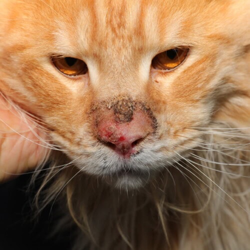
An allergic dermatosis can perfectly mimic feline cutaneous herpesvirus infection
Cowpox is rare but zoonotic, and it can be complicated by potentially fatal pulmonary involvement. It is confirmed by isolating the virus from cutaneous lesions, visualizing virions by electron microscopy, and identifying inclusion bodies by histology or cytology (McInerney et al., 2016).
Numerous infectious, allergic, immune-mediated, and neoplastic dermatoses affect the feline face, and their treatments as well as their prognoses differ. Indeed, none can be recognized by its morphology alone. The workup therefore begins with first-line examinations: skin scrapings to look for mites, cytology, fungal culture, and then, as indicated, antiparasitic trial, elimination diet, bacterial culture, and biopsies (Coyner, 2020).
Two anamnestic elements are more reliably orienting than lesion morphology. The first is lack of response to, or worsening with, corticosteroid therapy. The second is the existence of a reactivation trigger: corticosteroid therapy, stress, or communal living.
4.2 Cytology: An Orienting Examination
Cytology does not provide the diagnosis. Indeed, inclusion bodies, already difficult to find on histological sections, are practically impossible to observe on cytological smears. The sample is collected by impression smear from the ulcer or from the inner surface of the crust; it is constantly contaminated with blood and shows an eosinophilic exudate mixed with neutrophils and macrophages in variable proportions. This should by no means lead to a diagnosis of allergy, because an abundant eosinophilic infiltrate is also observed on the facial lesions of allergic cats, and the error could lead to systemic corticosteroid therapy whose consequences can be serious in an infected cat. Three elements combined should instead raise suspicion of a viral cause and prompt a biopsy: ulcers covered with hemorrhagic crusts, absence of pruritus, and a history of upper respiratory signs or conjunctivitis (Albanese, 2017, pp. 185–189).
4.3 Histopathology
Skin biopsy remains the first-line examination. Definitive histological diagnosis rests on the identification of slightly basophilic intranuclear inclusion bodies, with chromatin margination, in intact epithelial cells adjacent to necrotic areas (Persico et al., 2011).
The microscopic picture combines full-thickness epidermal necrosis extending to adnexa and dermis, and an eosinophilic and neutrophilic infiltrate of variable intensity (Persico et al., 2011). The authors of the original description emphasize three features that mimic eosinophilic granuloma complex: some adnexa are destroyed, free hairs are present in the dermis surrounded by numerous eosinophils, and collagen undergoes focal degeneration. The inclusion bodies fill the nucleus and are accompanied by chromatin margination and cytoplasmic swelling (Hargis et al., 1999). Other reports classify them as Cowdry type A inclusions (Suchy et al., 2000).
Three additional details complete this picture. The epidermis bordering the ulceration is thickened and spongiotic, and it is often here that the rare inclusions are found (Munday and Wilhelm, 2020). Multinucleated giant keratinocytes may be present in the surface and follicular epithelium, also bearing Cowdry type A inclusions, alongside suppurative folliculitis and furunculosis. The feature presented as specific to this dermatosis is necrosis of the epitrichial sweat glands (Miller et al., 2013). This is sought when inclusions are absent, bearing in mind that it is reported by this single 2013 source and does not appear in subsequent descriptions.
Three variants do not exclude this diagnosis. First, the infiltrate may be dominated not by eosinophils but by mast cells and plasma cells, with inclusions located in unidentifiable deep dermal cells on routine staining (Porcellato et al., 2018). Second, inclusions may be found in dermal macrophages rather than in the epithelium, with immunohistochemistry staining only macrophages in such cases (Sebastian Pineda et al., 2026a). Third, transmural vascular lesions, with fibrinoid necrosis and thrombosis of dermal arterioles and venules, were observed in six out of eight cases, possibly secondary to the extent of epidermal necrosis (Sebastian Pineda et al., 2026b).
4.4 Immunohistochemistry
Immunohistochemistry is the confirmatory examination of choice when clinical findings and histopathology suggest herpesvirus but inclusion bodies are absent (Persico et al., 2011). It demonstrates viral antigen in the nucleus and cytoplasm of epithelial cells located within lesions and in their immediate vicinity (Suchy et al., 2000). It is most useful when inclusions are scarce: it allowed two cases out of thirty to be attributed to herpesvirus in a population of eosinophilic dermatoses and stomatitis where routine histology had left them undetermined (Lee et al., 2010).
4.5 PCR and Electron Microscopy
The first study involved forty-seven biopsies. Viral DNA was detected in all nine samples from the five cats with herpetic dermatitis, in one out of seventeen samples from non-herpetic ulcerative dermatitis, and in none of the twenty-one samples from cats without lesions, yielding a sensitivity of 100% and a specificity of 95% using histology as the reference standard (Holland et al., 2006).
The second study, involving sixty-four biopsies, found twelve positive PCR results, only two of which were accompanied by a positive immunohistochemistry result; the authors attribute the excess of positives to amplification of vaccine-derived viral DNA, without being able to exclude amplification of latent virus, and conclude that PCR is suitable for initial screening but is insufficient for a definitive diagnosis (Persico et al., 2011).
A frequently overlooked source of false positives relates to sample collection itself: contact of the biopsy with conjunctival or nasal mucosa is sufficient to contaminate it (Miller et al., 2013), as is licking of the lesion by a carrier cat (Munday and Wilhelm, 2020). Viral isolation from a tissue fragment that has been surface-disinfected beforehand distinguishes active infection from simple contamination (Nuttall et al., 2019).
Three practical rules follow from this. Do not sample a cat recently vaccinated with a modified live vaccine (Thiry et al., 2009). Never confirm the diagnosis on a positive PCR result alone, which may be merely an incidental finding related to prior exposure (Nuttall et al., 2019). And do not dismiss the diagnosis on the basis of a negative PCR result either: in the original case series, one out of eight cats was PCR-negative while electron microscopy showed virions compatible with a herpesvirus (Hargis et al., 1999), whereas other authors consider amplification failure a reason for exclusion (Munday and Wilhelm, 2020).
Quantitative PCR provides a decisive addition. On formalin-fixed paraffin-embedded tissue, copy numbers of glycoprotein B and thymidine kinase genes are quantified (Mazzei et al., 2019).
Electron microscopy, available only in laboratories equipped for it, plays a supplementary role: it demonstrates in keratinocytes nucleocapsids of 100 to 125 nm whose morphology is compatible with a herpesvirus (Hargis et al., 1999), and it has served as a confirmatory method in atypical cases (Hargis et al., 1999; Porcellato et al., 2018).
Table 1 summarizes the confirmatory methods and their limitations.
Table 1 — Confirmatory methods for feline herpesvirus type 1 dermatitis
| Examination | What it demonstrates | Reported data | Main limitation |
|---|---|---|---|
| Histopathology (hematoxylin-eosin) | Epidermal, adnexal, and dermal necrosis; eosinophilic and neutrophilic infiltrate; intranuclear inclusion bodies | Inclusions found in all ten cases of the original series (Hargis et al., 1999) | Inclusions rare or absent in chronic or recurrent forms (Persico et al., 2011) |
| Immunohistochemistry | Viral antigen in the nucleus and cytoplasm of epithelial cells, sometimes in dermal macrophages | Two cases identified out of thirty eosinophilic dermatoses and stomatitis (Lee et al., 2010) | Not performed in all laboratories; requires prior clinical orientation |
| Conventional PCR | Viral DNA on fresh or paraffin-fixed tissue | Sensitivity 100%, specificity 95% vs. histology (Holland et al., 2006); twelve positives out of sixty-four, only two confirmed (Persico et al., 2011) | False positives from vaccine origin or latency; false negatives possible (Hargis et al., 1999) |
| Quantitative PCR | Copy number of glycoprotein B and thymidine kinase genes normalized to a housekeeping gene | Overexpression in all herpetic cases, absent in controls and non-herpetic dermatitis (Mazzei et al., 2019) | Limited availability; thresholds still laboratory-specific |
| Electron microscopy | Nucleocapsids of 100 to 125 nm in keratinocytes | Compatible virions in two cats, including one PCR-negative (Hargis et al., 1999) | Restricted access; requires appropriate fixation |
5 Treatment
5.1 Principles
Management rests on three components that must not be separated: a systemic antiviral active against feline herpesvirus, control of secondary bacterial infection, and above all elimination of reactivation triggers, foremost among which is corticosteroid therapy. No antiviral drug has authorization for the treatment of feline herpetic disease; their use therefore falls under extra-label prescribing, with the appropriate client disclosure that this entails (Thomasy and Maggs, 2016).
5.2 Famciclovir
Famciclovir is currently the systemic antiviral of choice in cats. It is a prodrug of penciclovir, a highly active molecule against feline herpesvirus, whereas famciclovir itself and its intermediate metabolite BRL42359 have no antiviral activity. The final conversion to penciclovir depends on hepatic aldehyde oxidase, whose activity in cats represents approximately 2% of that in humans, the lowest reported to date across all species studied (Thomasy and Maggs, 2016). This results in non-linear pharmacokinetics: doubling the dose does not double the plasma concentration of penciclovir; instead, absolute bioavailability falls from 67.1% at 15.6 mg/kg to 18.4% at 93.8 mg/kg (Qu et al., 2025).
Low doses are therefore ineffective: at 15 mg/kg of famciclovir every eight hours, the concentration remains well below that needed in vitro against the virus (Thomasy et al., 2007). The only blinded placebo-controlled trial used 90 mg/kg three times daily for twenty-one days in sixteen experimentally infected cats: clinical scores were reduced from day four to day eighteen, weight gain was sustained, and viral shedding was less frequent, with a maximum plasma penciclovir concentration of approximately 2 µg/mL (Thomasy et al., 2011). A retrospective study of fifty-nine cats with spontaneous disease showed a significantly shorter time to improvement and significantly greater improvement with 90 mg/kg than with 40 mg/kg, always three times daily (Thomasy et al., 2016).
Twice-daily administration is sufficient. Plasma and lacrimal penciclovir concentrations obtained with 90 mg/kg of famciclovir twice daily are comparable to those obtained with the same dose three times daily, whereas lower doses do not achieve sufficient lacrimal concentrations even when administered three times daily; the recommended regimen is therefore 90 mg/kg of famciclovir twice daily (Thomasy and Maggs, 2016). This regimen was retained in subsequent trials, including an experimental ocular infection study (Ledbetter et al., 2022) and a prospective randomized trial conducted in 373 kittens, in which the addition of famciclovir to doxycycline reduced the time to clinical cure in mild forms and, across the entire cohort, the frequency of corneal involvement (Vernau et al., 2024).
The duration of treatment is not standardized: in the retrospective series of fifty-nine cats, it ranged from four to 882 days, and one case of ulcerative facial dermatitis was controlled within thirty days (Thomasy et al., 2016). Duration is therefore governed by clinical progression rather than by a fixed protocol. Lower dosages still cited in some textbooks predate the pharmacokinetic studies; it therefore seems preferable to no longer follow them.
Dermatology-specific data remain limited. Four cats with herpesvirus-associated dermatitis all showed marked improvement with famciclovir, better than with previously tried strategies; three, however, subsequently relapsed. The doses used, 62.5 mg once or twice daily or 125 mg three times daily, correspond to posologies lower than those currently recommended (Malik et al., 2009). Four other cats in the retrospective series of fifty-nine animals had cutaneous involvement (Thomasy et al., 2016).
Tolerability is satisfactory, and markedly better than that of acyclovir and valacyclovir. Adverse effects attributable to famciclovir, most commonly gastrointestinal, were reported in 17% of cats receiving 40 or 90 mg/kg three times daily, with no difference between the two dose groups (Thomasy and Maggs, 2016). A complete blood count, biochemistry panel, and urinalysis should nonetheless be monitored in animals with concurrent disease or those intended for long-term treatment, and dosing intervals should be extended in cases of renal insufficiency (Thomasy and Maggs, 2016).
The pharmaceutical formulation matters, and this is often overlooked: most compounded preparations have proven unstable, with 400 mg/mL suspensions deviating by an average of 52.9% from the stated content (O’Leary et al., 2021). This variability in content may partly explain the limited efficacy of compounded famciclovir in a shelter-based trial (Mironovich et al., 2023). No functional resistance has been demonstrated to date (Lewin et al., 2023).
5.3 Agents to Avoid
Systemic acyclovir has no place in cats. The thymidine kinase of feline herpesvirus phosphorylates it far less efficiently than that of human herpes simplex virus, and oral administration of 50 mg/kg achieves only approximately one third of the necessary inhibitory concentration (Thomasy and Maggs, 2016); bone marrow aplasia and nephrotoxicity are additional concerns (Nuttall et al., 2019). Regarding topical application, one cat with presumed herpetic dermatitis responded to an acyclovir cream (Malik et al., 2009).
Valacyclovir is absolutely contraindicated. In experimentally infected cats, it caused potentially fatal hepatic and renal necrosis and bone marrow aplasia, without reducing either viral shedding or the severity of clinical signs, due to the toxic acyclovir concentrations reached (Thomasy and Maggs, 2016).
Foscarnet, finally, has only 8% oral bioavailability in cats and low activity against the virus; its use is not recommended (Thomasy and Maggs, 2016).
Three agents have feline data, all in ophthalmology. Ganciclovir 0.15% gel, three times daily, equals oral famciclovir at 90 mg/kg twice daily on clinical scores and corneal infiltrate (Ledbetter et al., 2022). Cidofovir 0.5% solution significantly reduces ocular viral load (Mironovich et al., 2023). Human 1% penciclovir dermatological cream, applied to the eye, is well tolerated and maintains lacrimal concentrations above the minimum inhibitory concentration for more than eight hours (Pe’er et al., 2025).
It is tempting to extrapolate these results to the skin, especially since 1% penciclovir cream is precisely a dermatological formulation. No study has done so in cats, but its use is described: creams intended for human cold sores, particularly those containing penciclovir, are presented as potentially useful and combinable with systemic famciclovir (Munday and Wilhelm, 2020). Topical imiquimod, applied two to three consecutive days per week, is also listed among the options mentioned (Miller et al., 2013).
5.5 Interferon
Recombinant feline omega interferon reduces in vitro the number and size of viral lysis plaques, and its effect at high concentrations exceeds that of human alpha-2b interferon (Siebeck et al., 2006).
Translation to clinical use is less convincing, except in dermatology where a detailed case report exists. A cat with confirmed herpetic facial dermatitis was successfully treated with omega interferon subcutaneously on days 0, 2, and 4, followed by a second series twelve weeks later, this time as perilesional injections given the marked local improvement obtained; cutaneous PCR remained positive after treatment, a finding whose significance is unknown (Gutzwiller et al., 2007). Dosages reported in textbooks vary: 1.5 MU/kg perilesionally and subcutaneously for omega interferon, 1 MU/m² subcutaneously three times weekly or 0.01 to 1 MU/kg once daily for up to three weeks for alpha interferon (Miller et al., 2013; Munday and Wilhelm, 2020).
Of the published clinical trials, only two were placebo-controlled and neither showed a significant effect (Thomasy and Maggs, 2016), and the authors who publish these protocols accompany them with explicit reservations. No author positions interferon within the therapeutic strategy. It should therefore be reserved for situations where famciclovir is poorly tolerated or ineffective, which is a matter of clinical judgment rather than an established recommendation.
5.6 L-Lysine
Six studies have examined lysine in cats, all published before 2015, and no study has been published since.
A systematic review of seventeen publications concludes as follows: lysine has no inherent antiviral properties, the arginine antagonism on which its supposed mechanism is based is not observed in cats, it does not modify viral replication in vitro, and available clinical trials have shown no efficacy, with some even reporting higher infection frequency and increased severity (Bol and Bunnik, 2015). Other authors draw from the same studies the conclusion that the product is safe when given orally and that bolus administration might reduce viral shedding in latently infected cats and signs during primary exposure (Thomasy and Maggs, 2016). These results come from experimental studies conducted in small numbers of inoculated cats, whereas the only study conducted in naturally infected cats — 144 shelter animals receiving 250 to 500 mg per day — demonstrated no effect or benefit. The same authors also point out that no trial has ever been conducted in the population in which the molecule is actually prescribed.
Dietary supplementation refers not to oral administration in general, but to lysine incorporated into the feed and ingested at mealtimes, as opposed to bolus tablets or paste given directly to the animal. This enriched diet proved detrimental: disease was more severe and viral shedding was greater than on a basal diet. The authors explain this as follows: a sick cat eats less, so its lysine intake falls precisely at the time when it might theoretically benefit from it (Thomasy and Maggs, 2016). Lowering arginine also risks potentially fatal hyperammonemia in a species unable to synthesize this amino acid (Bol and Bunnik, 2015).
Lysine therefore has no place in the treatment of herpetic dermatitis.
5.7 Corticosteroids and Immunomodulators
Systemic corticosteroid therapy must be avoided as soon as the diagnosis is being considered. Eight out of ten cats showed no improvement or worsened with corticosteroids (Hargis et al., 1999), and worsening after corticosteroids is also reported in atypical forms (Porcellato et al., 2018). Beyond their ineffectiveness, corticosteroids are among the recognized causes of viral reactivation and shedding (Thiry et al., 2009). When faced with a feline ulcerative dermatitis that does not respond to corticosteroids, the appropriate course of action is to perform a biopsy rather than increase the dose. As a general principle, corticosteroids and ciclosporin should not be administered to a cat with viral dermatitis (Nuttall et al., 2019).
5.8 Adjunctive Treatments
Supportive care is part of the treatment, precisely because specific antivirals may prove ineffective (Nuttall et al., 2019). Correcting the triggering factor, discontinuing all immunosuppressive treatment, feeding the animal properly, and reducing its stress are sometimes sufficient to achieve recovery (Miller et al., 2013). Secondary bacterial infection is constant in extensive ulcerative forms and justifies antibiotic therapy, ideally guided by culture (Flacke et al., 2015).
Surgical excision retains a place for small, well-demarcated lesions. Indeed, two lesions healed solely as a result of the biopsy procedure itself, and a residual lesion after medical treatment can similarly be resected (Hargis et al., 1999).
Table 2 presents the available agents, their level of evidence, and their dosages.
Table 2 — Agents used against feline herpesvirus type 1 and level of evidence in cats
| Agent | Level of evidence in cats | Documented dosage | Remarks |
|---|---|---|---|
| Famciclovir | Placebo-controlled trial on experimental infection (Thomasy et al., 2011); retrospective series of 59 cases including 4 dermatitis cases (Thomasy et al., 2016) | 90 mg/kg twice daily orally (Thomasy and Maggs, 2016) | Reference systemic antiviral; non-linear pharmacokinetics; gastrointestinal adverse effects in 17% of animals |
| Topical ganciclovir | Randomized placebo-controlled trial, ocular route (Ledbetter et al., 2022) | 0.15% ophthalmic gel, three times daily | Efficacy comparable to oral famciclovir on the eye; cutaneous use not studied |
| Topical cidofovir | Controlled shelter trial, ocular route (Mironovich et al., 2023) | 0.5% ophthalmic solution, twice daily | Significant reduction in ocular viral load |
| 1% penciclovir cream | Lacrimal tolerance and pharmacokinetics (Pe’er et al., 2025); cutaneous use reported from personal communication (Munday and Wilhelm, 2020) | Two applications per day | Human dermatological formulation, combinable with oral famciclovir |
| Recombinant feline omega interferon | In vitro data (Siebeck et al., 2006); one dermatitis case successfully treated (Gutzwiller et al., 2007) | 1.5 MU/kg perilesionally and subcutaneously, expert opinion from 2013 (Miller et al., 2013) | Second-line option; controlled trials inconclusive (Thomasy and Maggs, 2016) |
| L-lysine | Systematic review of seventeen publications (Bol and Bunnik, 2015); nuanced reading of the same studies (Thomasy and Maggs, 2016) | Not applicable | No demonstrated benefit in cats with recurrences; dietary supplementation detrimental |
6 Progression, Prognosis, and Prevention
6.1 Progression and Recurrences
The course is chronic and unpredictable. Lesions are often progressive and persistent, and healing may leave a scar (Hargis et al., 1999). Furthermore, recurrences are common, even after a clear initial response: three of the four cats improved with famciclovir relapsed (Malik et al., 2009), and a recurrence occurred seven months after the initial biopsy in an atypical limb form (Sebastian Pineda et al., 2026a). Affected cats often become persistent carriers, and any immunosuppression or stress can trigger a recurrence (Nuttall et al., 2019). Conversely, dermatitis may resolve spontaneously, but the frequency of such spontaneous recoveries remains unknown due to the lack of reported untreated cats (Munday and Wilhelm, 2020); these remissions may falsely suggest that a treatment has been effective.
6.2 Vaccination
Vaccination provides reasonable protection against disease but not against infection, even if viral shedding may be reduced (Gaskell et al., 2007). The recommended protocol consists of two injections, at nine and twelve weeks of age, followed by a first booster one year later. Subsequent boosters are annual in cats with exposure risk, and spaced three years apart in low-risk cats, such as strictly indoor cats. Since a recovered cat is generally not protected for life against new episodes, vaccination is also recommended in these animals (Thiry et al., 2009).
A cat recently vaccinated with a modified live vaccine should not be sampled for PCR, at the risk of obtaining a non-significant positive result (Thiry et al., 2009), with vaccine-derived DNA being precisely the explanation proposed for cutaneous false positives (Persico et al., 2011).
The possible role of the vaccine in triggering lesions remains debated. The original case series found no association between recent vaccination, including by the intranasal route, and the appearance of lesions (Hargis et al., 1999). In contrast, in the two cases of flank and thigh dermatitis, modified live vaccination near the lesional site preceded lesions by a few weeks in one cat and by a few days in the other, in whom lesions developed at the injection site with a histological appearance distinct from classic injection site reactions (Sánchez et al., 2012).
6.3 Environmental Control
To prevent recurrences, the circumstances that reactivate the virus must be reduced, with stress and corticosteroid therapy being the two recognized causes (Thiry et al., 2009). Four measures follow directly from what is known about the viral cycle, without their effect being quantified: reduce animal density, isolate shedding sick animals, apply basic hygiene measures, and avoid all unnecessary corticosteroid therapy. The virus, which is fragile, is susceptible to most disinfectants, antiseptics, and detergents (Thiry et al., 2009).
Conclusion
Feline cutaneous herpesvirus infection is a well-recognized diagnostic pitfall. Its ulcerative and crusted presentation, most often facial but sometimes elsewhere, and its eosinophilic infiltrate can easily mimic allergic dermatitis or an eosinophilic granuloma complex lesion. The scarcity of inclusion bodies further obscures the picture. When in doubt, biopsy the margin of any feline facial ulcerative lesion that fails to respond to corticosteroids rather than increasing the dose, and explicitly request that the laboratory search for herpesvirus, bearing in mind that a negative immunohistochemistry result is not definitive and that an isolated positive PCR result is insufficient.
References
Albanese F. Canine and Feline Skin Cytology. A Comprehensive and Illustrated Guide to the Interpretation of Skin Lesions via Cytological Examination. Cham: Springer International Publishing; 2017. pp. 61–62, 185–189.
Bol S, Bunnik EM. Lysine supplementation is not effective for the prevention or treatment of feline herpesvirus 1 infection in cats: a systematic review. BMC Vet Res. 2015;11:284.
Coyner K. Distinguishing between dermatologic disorders of the face, nasal planum, and ears: great lookalikes in feline dermatology. Vet Clin North Am Small Anim Pract. 2020;50(4):823–882.
De Lucia M, Cabré M, Denti D, Mezzalira G, Rondena M, Furlanello T. Presumptive herpesvirus-associated erythema multiforme in a cat. Vet Dermatol. 2021;32(1):86–e16.
Flacke GL, Schmidt-Küntzel A, Marker L. Treatment of chronic herpesviral dermatitis in a captive cheetah (Acinonyx jubatus) in Namibia. J Zoo Wildl Med. 2015;46(3):641–646.
Gaskell R, Dawson S, Radford A, Thiry E. Feline herpesvirus. Vet Res. 2007;38(2):337–354.
Gutzwiller ME, Brachelente C, Taglinger K, Suter MM, Weissenböck H, Roosje PJ. Feline herpes dermatitis treated with interferon omega. Vet Dermatol. 2007;18(1):50–54.
Hargis AM, Ginn PE, Mansell JEKL, Garber RL. Ulcerative facial and nasal dermatitis and stomatitis in cats associated with feline herpesvirus 1. Vet Dermatol. 1999;10(4):267–274.
Hargis AM, Ginn PE. Feline herpesvirus 1-associated facial and nasal dermatitis and stomatitis in domestic cats. Vet Clin North Am Small Anim Pract. 1999;29(6):1281–1290.
Holland JL, Outerbridge CA, Affolter VK, Maggs DJ. Detection of feline herpesvirus 1 DNA in skin biopsy specimens from cats with or without dermatitis. J Am Vet Med Assoc. 2006;229(9):1442–1446.
Lappin MR, Roycroft LM. Effect of ciclosporin and methylprednisolone acetate on cats previously infected with feline herpesvirus 1. J Feline Med Surg. 2015;17(4):353–358.
Ledbetter EC, Badanes ZI, Chan RX, Donohue LK, Hayot NL, Harman RM, Van de Walle GR, Mohammed HO. Comparative efficacy of topical ophthalmic ganciclovir and oral famciclovir in cats with experimental ocular feline herpesvirus-1 epithelial infection. J Ocul Pharmacol Ther. 2022;38(5):339–347.
Lee M, Bosward KL, Norris JM. Immunohistological evaluation of feline herpesvirus-1 infection in feline eosinophilic dermatoses or stomatitis. J Feline Med Surg. 2010;12(2):72–79.
Lewin AC, Ineck NE, Mironovich MA, Marino ME, Liu CC, Emelogu U, Mills EP, Camacho-Luna P, Carter RT. Surveillance for feline herpesvirus type 1 mutation and development of resistance in cats treated with antiviral medications. Front Vet Sci. 2023;10:1197249.
Malik R, Lessels NS, Webb S, Meek M, Graham PG, Vitale C, Norris JM, Power H. Treatment of feline herpesvirus-1 associated disease in cats with famciclovir and related drugs. J Feline Med Surg. 2009;11(1):40–48.
Mazzei M, Vascellari M, Zanardello C, Melchiotti E, Vannini S, Forzan M, Marchetti V, Albanese F, Abramo F. Quantitative real time polymerase chain reaction (qRT-PCR) and RNAscope in situ hybridization (RNA-ISH) as effective tools to diagnose feline herpesvirus-1-associated dermatitis. Vet Dermatol. 2019;30(6):491–e147.
McInerney J, Papasouliotis K, Simpson K, English K, Cook S, Milne E, Gunn-Moore DA. Pulmonary cowpox in cats: five cases. J Feline Med Surg. 2016;18(6):518–525.
Miller WH, Griffin CE, Campbell KL. Viral, rickettsial, and protozoal skin diseases. In: Muller and Kirk’s Small Animal Dermatology. 7th ed. St. Louis: Elsevier Mosby; 2013. pp. 343–362.
Mironovich MA, Yoon A, Marino ME, Ineck NE, Liu CC, Carter RT, Lewin AC. Evaluation of compounded cidofovir, famciclovir, and ganciclovir for the treatment of feline herpesvirus ocular surface disease in shelter-housed cats. Vet Ophthalmol. 2023;26(Suppl 1):143–153.
Munday JS, Wilhelm S. Viral diseases. In: Noli C, Colombo S, eds. Feline Dermatology. Cham: Springer Nature Switzerland; 2020. pp. 359–380.
Nuttall T, Eisenschenk M, Heinrich NA, Harvey RG. Feline cutaneous herpesvirus and calicivirus infections. In: Skin Diseases of the Dog and Cat. 3rd ed. Boca Raton: CRC Press; 2019. pp. 159–160.
O’Leary LM, Allbaugh RA, Schrunk DE, Olsen TE, Sebbag L. Variable accuracy, precision, and consistency of compounded famciclovir formulated for management of feline herpesvirus-1 in cats. Vet Ophthalmol. 2021;24(6):627–638.
Pe’er O, Thomasy SM, Feuer E, Knych HK, Ofri R, Sebbag L. Ocular tolerance and tear film pharmacokinetics of 1% penciclovir cream in cats. Res Vet Sci. 2025;196:105909.
Persico P, Roccabianca P, Corona A, Vercelli A, Cornegliani L. Detection of feline herpes virus 1 via polymerase chain reaction and immunohistochemistry in cats with ulcerative facial dermatitis, eosinophilic granuloma complex reaction patterns and mosquito bite hypersensitivity. Vet Dermatol. 2011;22(6):521–527.
Porcellato I, Luciani L, Marenzoni ML, Santagostino SF, Sforna M, Mechelli L, Brachelente C. Feline herpesvirus ulcerative dermatitis: an atypical case? Vet Dermatol. 2018;29(3):258–e96.
Qu M, Ke X, Zhang Z, Zhang L. Non-linear pharmacokinetics of penciclovir in healthy cats after single and multiple oral administration of famciclovir. Front Vet Sci. 2025;12:1695827.
Roberts ES, VanLare KA, Roycroft LM, King S. Effect of high-dose ciclosporin on the immune response to primary and booster vaccination in immunocompetent cats. J Feline Med Surg. 2015;17(2):101–109.
Sebastian Pineda DA, Maboni G, Garcia G, Baker C, Bacon L, Rissi DR. Atypical felid alphaherpesvirus 1 dermatitis in a cat. J Vet Diagn Invest. 2026a;38(2):330–334.
Sebastian Pineda DA, Schneider SM, Mendes RE, Rissi DR. Dermal inflammatory vascular changes associated with felid alphaherpesvirus 1 dermatitis in cats. J Comp Pathol. 2026b;227:61–64.
Siebeck N, Hurley DJ, Garcia M, Greene CE, Köstlin RG, Moore PA, Dietrich UM. Effects of human recombinant alpha-2b interferon and feline recombinant omega interferon on in vitro replication of feline herpesvirus-1. Am J Vet Res. 2006;67(8):1406–1411.
Suchy A, Bauder B, Gelbmann W, Löhr CV, Teifke JP, Weissenböck H. Diagnosis of feline herpesvirus infection by immunohistochemistry, polymerase chain reaction, and in situ hybridization. J Vet Diagn Invest. 2000;12(2):186–191.
Synowiec A, Dąbrowska A, Pachota M, Baouche M, Owczarek K, Niżański W, Pyrc K. Feline herpesvirus 1 (FHV-1) enters the cell by receptor-mediated endocytosis. J Virol. 2023;97(8):e0068123.
Sánchez MD, Goldschmidt MH, Mauldin EA. Herpesvirus dermatitis in two cats without facial lesions. Vet Dermatol. 2012;23(2):171–173, e35.
Thiry E, Addie D, Belák S, Boucraut-Baralon C, Egberink H, Frymus T, Gruffydd-Jones T, Hartmann K, Hosie MJ, Lloret A, Lutz H, Marsilio F, Pennisi MG, Radford AD, Truyen U, Horzinek MC. Feline herpesvirus infection. ABCD guidelines on prevention and management. J Feline Med Surg. 2009;11(7):547–555.
Thomasy SM, Lim CC, Reilly CM, Kass PH, Lappin MR, Maggs DJ. Evaluation of orally administered famciclovir in cats experimentally infected with feline herpesvirus type-1. Am J Vet Res. 2011;72(1):85–95.
Thomasy SM, Maggs DJ, Moulin NK, Stanley SD. Pharmacokinetics and safety of penciclovir following oral administration of famciclovir to cats. Am J Vet Res. 2007;68(11):1252–1258.
Thomasy SM, Maggs DJ. A review of antiviral drugs and other compounds with activity against feline herpesvirus type 1. Vet Ophthalmol. 2016;19(Suppl 1):119–130.
Thomasy SM, Shull O, Outerbridge CA, Lim CC, Freeman KS, Strom AR, Kass PH, Maggs DJ. Oral administration of famciclovir for treatment of spontaneous ocular, respiratory, or dermatologic disease attributed to feline herpesvirus type 1: 59 cases (2006–2013). J Am Vet Med Assoc. 2016;249(5):526–538.
Vernau KM, Kim S, Thomasy SM, Lucyshyn DR, Purpura J, Montgomery E, Surmick JD, Dubelko AR, Moussavi A, Kass PH, Maggs DJ. Doxycycline with or without famciclovir for infectious ophthalmic and respiratory disease: a prospective, randomized, masked, placebo-controlled trial in 373 kittens. J Feline Med Surg. 2024;26(11):1098612X241278413.