


Dermatophytosis is a superficial infection of the skin and hair coat caused by keratinophilic fungi of the genera Microsporum, Trichophyton and Nannizzia. In dogs and cats, Microsporum canis is by far the dominant agent. The disease is contagious, infectious and transmissible to people, which gives it a place of its own in the dermatology consultation. The practitioner treats an animal and protects a household.
Roughly half of the people exposed to an infected cat, whether or not that cat shows signs, will contract the infection, and in 30 to 70% of households harboring an infected cat at least one person is affected (Miller et al., 2013). Treatment itself has changed. Not in its principles, which are set by international consensus guidelines, but in the means available: products have come and gone from the market, and a resistance mechanism has emerged. This is why 2026 was the right moment for a full review of both the drugs that remain available and the protocols as they now stand. Availability differs on either side of the Atlantic, and this review states for each drug what is approved in the United States and what is authorized in Europe, since the published evidence was generated on both.
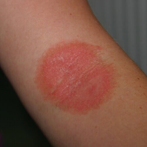
Dermatophytosis in a human
1 Developments since the reference protocols
1.1 Why treat a disease that resolves on its own?
Dermatophytosis is not life-threatening and resolves without intervention. If we treat, it is to shorten the course of the disease and limit transmission (Moriello, 2019a).
Five dogs aged 6 to 18 months with generalized dermatophytosis received an inert substance given orally once daily with food. Signs resolved in 3 of them after 4 to 8 weeks. The other 2, which had not improved, recovered after 6 weeks of ketoconazole (Medleau et al., 1992). In cats, the disease is described as self-limiting in a large proportion of cases, with hair loss and scaling as the only manifestations; multifocal or generalized forms are seen mainly in kittens and immunocompromised animals (Frymus et al., 2013).
Cure rates in control groups are therefore not artifacts. The 6 of 40 cats that recovered on placebo in the pivotal itraconazole trial illustrate this natural course. It is against that background that the effect of any treatment must be measured. And since the animal would recover in any case, the therapeutic goal is to shorten the contagious period and clear the environment.
1.2 The 2017 consensus guidelines and what they established
The reference document for the discipline remains the clinical consensus guidelines of the World Association for Veterinary Dermatology, published in 2017 (Moriello et al., 2017).
No diagnostic test is designated there as a gold standard. Wood’s lamp examination and direct examination of hairs both have good predictive value, positive as well as negative.
As for treatment, cure requires the simultaneous use of an oral systemic antifungal and a topical antifungal applied to the coat. Since the animal carries infective spores on hairs that are not all visibly affected, neither route is sufficient on its own. Systemic antifungals are further described as having a wide margin of safety, and physical cleaning together with application of an antifungal in the environment is described as essential to decontamination of the premises. Finally, the authors point out that serious complications of transmission to humans are exceptional.
1.3 Product availability in the United States and in Europe
Several of the products cited in older international guidelines cannot be obtained everywhere, and what is on the shelf now differs sharply between the two continents.
In the United States, itraconazole is the only systemic antifungal carrying an approval in this indication. Ketoconazole holds a human approval but none as a veterinary drug, so its use in dogs is extra-label. Griseofulvin occupies an ambiguous position: it is described as the only approved systemic antifungal for veterinary use, that approval covering the horse, while use in dogs and cats is extra-label and the presentations formerly approved for dogs are no longer marketed. Lime sulfur is available, but as an unapproved drug: its labels carry the statement that the agency has not found it safe and effective and has not approved the labeling. The compound is registered with the Environmental Protection Agency, for agricultural uses and for topical application against livestock mites. Supply has also become uneven, since some manufacturers discontinued their lime sulfur products after their raw material supplier withdrew from the pet market following a change in environmental labeling guidance, without the general availability of the compound being affected.
In Europe the picture is reversed on two counts. Enilconazole, which is not marketed in the United States (VCA, 2026), is available there as a 100 mg/ml concentrate for cutaneous spray emulsion, IMAVERAL (Audevard), authorized in dogs, cats, cattle and equids for dermatophytosis caused by Microsporum canis, Microsporum gypseum, Trichophyton verrucosum, Trichophyton mentagrophytes and Trichophyton equinum (ANSES-ANMV, 2026). It is the only whole-body topical with established residual activity that European practitioners can obtain, and it may be applied in the environment at a concentration five times higher than that used on animals. Ketoconazole holds a veterinary authorization in dogs there, and griseofulvin in dogs and cats. Conversely, lime sulfur, which underpins the entire North American literature, corresponds to no authorized veterinary medicine in the European Union.
Two products that structured older protocols have disappeared from the French market. The veterinary griseofulvin tablet, FULVIDERM (Virbac), still holds a valid marketing authorization in the European veterinary medicines register but can no longer be ordered, leaving a single oral powder, DERMOGINE (Dopharma France), at 100 mg of griseofulvin per gram, authorized in dogs, cats and equids (ANSES-ANMV, 2026). The enilconazole smoke generator, CLINAFARM (Elanco France), long the reference tool in catteries and shelters, is no longer marketed in France; regulated as a biocide rather than as a veterinary medicine, it leaves a gap that nothing strictly equivalent fills, and its withdrawal makes part of the cattery protocols written before it obsolete. In human medicine, Sanofi discontinued GRISEFULINE 250 mg and 500 mg in July 2021, because of manufacturing difficulties that could not be resolved (ANSM, 2021).
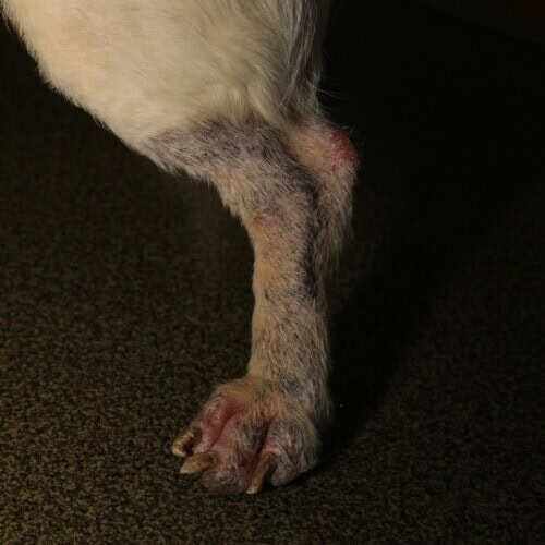
Extensive dermatophytosis in a dog
1.4 The emergence of allylamine-resistant dermatophytes
The most striking new development since 2013 concerns not a drug but a fungus. Trichophyton indotineae, first described as genotype VIII of the Trichophyton mentagrophytes complex, emerged from the Indian subcontinent and has spread internationally (Ahmad, 2026).
Point mutations in the squalene epoxidase gene alter the target of terbinafine. Three substitutions have been named: Leu393Phe, Phe397Leu and Ala448Thr. These mutations confer high minimum inhibitory concentrations for terbinafine, and they have been detected in isolates of both human and animal origin, which points to the circulation of stable, transmissible resistant clones (Ahmad, 2026).
Whole-genome sequencing of 13 Indian isolates, 9 of human and 4 of canine origin, showed few polymorphic differences between the two origins, which argues for zoonotic transmission and rapid spread over long distances (Thakur et al., 2025). That said, two canine strains from northern India characterized elsewhere proved susceptible to terbinafine, with no substitution at positions 393 or 397 (Thomas et al., 2025). Resistance therefore does not travel with the species, it travels with certain clones.
In Europe, two T. mentagrophytes isolates from Polish hedgehogs carried the Leu393Phe substitution, with a minimum inhibitory concentration of 2 µg/ml for terbinafine (Gnat et al., 2021).
Microsporum canis, the agent of nearly all canine and feline cases, is also capable of resistance, but by another route. The first terbinafine-resistant strain isolated from a cat was reported in 2018: a two-year-old exotic shorthair with alopecia and mild scaling of the trunk, whose strain showed a minimum inhibitory concentration above 32 µg/ml for terbinafine and of 0.023 µg/ml for itraconazole. The cat recovered on systemic itraconazole (Hsiao et al., 2018). The mechanism involved is not a squalene epoxidase mutation but overexpression of ABC transporters: in a resistant strain, expression of the PDR1, MDR1, MDR2 and MDR4 genes was two to four times higher than in susceptible strains, and adding the efflux blocker FK506 lowered the minimum inhibitory concentration from more than 32 to 8 µg/ml (Kano et al., 2018). Efflux-mediated resistance therefore does not necessarily spare the azoles, unlike a mutation of the terbinafine target.
Its frequency, however, remains low. Among 348 M. canis isolates from mainland China studied over 27 years using the M38-A3 method of the Clinical and Laboratory Standards Institute, in a human series that included 54 cases of tinea capitis in children, two strains proved resistant to griseofulvin with a minimum inhibitory concentration of 64 µg/ml, one to fluconazole at the same value, and two to terbinafine at 16 µg/ml. The great majority of strains retained low minimum inhibitory concentrations for the 11 drugs tested, and analysis of their distribution revealed no clear resistance pattern (Liang et al., 2025). In Central Asia, among human clinical isolates, all 57 M. canis strains tested by the EUCAST method remained susceptible to terbinafine, whereas 4 of 33 Trichophyton strains were resistant (Aimoldina et al., 2026). These two series cannot be compared point for point: minimum inhibitory concentration values produced by the American and the European methods are not superimposable, and only the order of magnitude of the resistance frequency carries across from one study to the other.
Resistance in M. canis therefore exists, but it is rare, and it arises by a mechanism distinct from that of T. indotineae. Moreover, no clinical series to date links squalene epoxidase mutations to treatment failure in dogs or cats. The immediate consequence is therefore diagnostic before it is therapeutic: it bears on species identification in cases of failure, not on first-line drug choice. It is all the more demanding because identification by culture does not distinguish T. indotineae from the other Trichophyton species: that requires sequencing of the ITS region completed by sequencing of the HMG gene, which encodes an HMG-box transcription factor whose sequence separates species that the ITS region alone does not, or mass spectrometry with an up-to-date library, or sequencing of the squalene epoxidase gene (Ahmad, 2026).
2 Pharmacology of the usable antifungals
2.1 Azoles: mechanism, spectrum and activity in keratin
Azoles inhibit 14-alpha-demethylase, the fungal cytochrome P450 enzyme that converts lanosterol into ergosterol. The fungal membrane is then deprived of its constituent sterol and enriched in methylated precursors. Its permeability is disrupted and fungal growth stops. The effect is fungistatic at the concentrations achieved clinically.
This inhibition is not perfectly selective, however, and that is where the adverse effects of the class come from. The same cytochrome P450 enzymes are involved in mammalian steroidogenesis and in the hepatic metabolism of many drugs, hence the hormonal disturbances and drug interactions observed, especially with ketoconazole.
Itraconazole accumulates in hair and skin, where it persists above the therapeutic threshold during the off weeks of treatment, and beyond the last dose given. It is this tissue persistence, and it alone, that makes the alternating-week regimen possible.
2.2 Allylamines: fungicidal activity and persistence
Terbinafine acts further upstream in the same pathway. It inhibits squalene epoxidase, the fungal enzyme that converts squalene into squalene epoxide. Two consequences add up: ergosterol is no longer produced, and squalene accumulates in the fungal cell to toxic levels. That accumulation kills the fungus rather than merely preventing it from growing. Terbinafine is therefore fungicidal against dermatophytes, where the azoles are fungistatic.
It is this same enzyme that the mutations described above alter, which explains why resistance acquired by target mutation affects terbinafine and spares the azoles. That reservation does not apply to efflux-mediated resistance, which exports several drug classes indiscriminately and therefore does not necessarily spare the azoles.
2.3 Griseofulvin: residual place and constraints
Griseofulvin is a fungistatic antibiotic produced by Penicillium griseofulvum. It binds to the intracellular microtubules of the dermatophyte and blocks their polymerization, arresting mitosis in metaphase. It reaches the cells that synthesize keratin precursors and becomes associated with keratin in the skin appendages. Its metabolism is essentially hepatic and its elimination mainly urinary. Its spectrum is limited to dermatophytes, chiefly Microsporum spp. and Trichophyton spp. (ANSES-ANMV, 2026).
Its intestinal absorption is poor and depends on the lipid content of the meal, which means it must be given with fatty food. Its safety constraints, discussed below, have relegated it behind the azoles wherever those are available.
3 Systemic treatment
3.1 Itraconazole
Itraconazole is today the first-line drug in the cat. The European feline guidelines already gave it that place (Frymus et al., 2013), and subsequent data have left it there.
The 10 mg/ml oral solution for cats, ITRAFUNGOL, was approved by the Food and Drug Administration on November 10, 2016 for the treatment of dermatophytosis caused by Microsporum canis in cats, and it remains the only systemic treatment for dermatophytosis approved in that country (FDA, 2016). It is given at 5 mg/kg once daily, that is 0.5 ml/kg, on alternating weeks, for three treatment cycles: dosing in weeks 1, 3 and 5, with no dosing in weeks 2 and 4. The same product holds a French authorization delivered in 2004, with an identical regimen (ANSES-ANMV, 2026).
Use calls for caution in cats with renal dysfunction or impaired liver function, and treatment is stopped if signs suggestive of liver dysfunction appear. Reproductive safety is not established: in a study of 16 queens given the oral solution at 5 mg/kg for a total of 21 days during gestation or lactation, there was a high frequency of partial and total fetal resorption, abnormal fetuses and abnormal maternal behavior; concurrent infectious disease in some of the cats made a definitive causal relationship difficult to establish, but the results do not support safe use in pregnant queens (FDA, 2016). Two interactions have been observed in cats: combination with cefovecin caused vomiting and hepatic and renal disturbances, and combination with tolfenamic acid caused motor incoordination, fecal retention and dehydration. To these are added the interactions linked to cytochrome P450 3A4 and to P-glycoproteins, described in human medicine, which increase plasma concentrations of midazolam, cyclosporine, digoxin, chloramphenicol, ivermectin and methylprednisolone (ANSES-ANMV, 2026).
The label states that cats treated with itraconazole may continue to contaminate other animals, dogs included, until mycological cure is achieved. The pivotal study enrolled 80 cats infected with M. canis, allocated to itraconazole solution or placebo, with no associated topical treatment. Cure on Wood’s lamp examination, defined as the absence of fluorescence at the base and mid-shaft of the hair, was achieved in 39 of 40 treated cats versus 6 of 40 controls, and time to mycological cure was significantly shorter in the treated group. In the two cats that failed, itraconazole minimum inhibitory concentrations remained within the susceptible range, which rules out resistance as the explanation. Field safety was assessed in 266 cats: 13% showed one or more elevated liver enzymes and 3% gastrointestinal upset, namely decreased appetite, vomiting or diarrhea (FDA, 2016).
The review of published studies sets out the expected timeframes: itraconazole at 10 mg/kg once daily, or on the combined continuous-then-pulse regimen, cured infected animals in 56 to 70 days, whereas low-dose protocols of 1.5 to 3 mg/kg in 15-day cycles required one to three cycles, that is 15 to 45 days (Moriello, 2004).
Earlier data under natural conditions had already established the efficacy of the drug, at lower doses and with poorer tolerance. Among 15 cats with M. canis dermatophytosis treated with oral itraconazole at 1.5 to 3 mg/kg once daily for 15 days, 8 recovered completely, 6 of them after a single course. Five cats vomited or became anorectic at the higher doses, which required the dose to be reduced progressively. Six of the 15 animals had previously failed griseofulvin at 10 mg/kg for 60 days (Mancianti et al., 1998).
The combined continuous-then-pulse regimen was evaluated in 9 cats: itraconazole at 10 mg/kg once daily for 28 days, then on alternating weeks at the same dose, stopping after two consecutive negative cultures. Eight of the 9 cats were cured at 56 days (Colombo et al., 2001). The sample is small and the authors themselves present these results as preliminary, pending a controlled study that never followed.
The approval does not cover the dog, in which no controlled trial has evaluated itraconazole alone, so canine use is extra-label. A retrospective study of 64 dogs infected with the Trichophyton mentagrophytes complex in the central United States over the period 1997 to 2020 concludes that ketoconazole, itraconazole and terbinafine appear equivalent as systemic options in this indication (Pieper et al., 2023). The level of evidence is that of a retrospective series, but it is the most substantial canine data available.
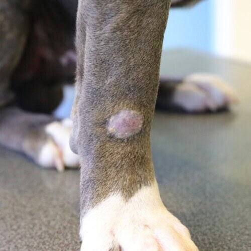
Few licensed systemic treatments exist in the dog
One practical point deserves emphasis, because it decides whether the drug works at all. Both the extent and the rate of itraconazole absorption vary widely between preparations compounded by different pharmacies, by the same pharmacy at different times, and under different manufacturing conditions. The approved formulations owe their bioavailability to specific excipients, a solubilizing cyclodextrin in the oral solution and coated drug spheres in the capsules, which compounded product made from bulk chemical lacks, resulting in very poor gastrointestinal absorption. The agency accordingly recommends prescribing the approved product rather than itraconazole compounded from bulk drug substances (FDA, 2016).
3.2 Terbinafine
Terbinafine holds no veterinary approval on either continent. In the United States it is prescribed extra-label under the Animal Medicinal Drug Use Clarification Act, which permits use of a human-approved drug in animals within a valid veterinarian-client-patient relationship. In Europe the equivalent mechanism, the cascade, is more restrictive, since it opens only when no authorized medicine is suitable for the species and indication: ketoconazole being authorized in dogs there, and griseofulvin in dogs and cats, the absence of an authorized alternative cannot be the justification. The place of terbinafine has nonetheless become established, for two reasons: its fungicidal activity, and its availability where itraconazole is expensive or lacking.
The strongest data come from a shelter study of 85 cats with naturally occurring M. canis dermatophytosis, monitored by weekly toothbrush culture with colony-forming unit counts. The toothbrush technique consists of methodically combing the entire coat with a new sterile toothbrush, then inoculating the brush itself onto the culture medium: it collects infected hairs from the whole body, including from outside visible lesions, which makes it superior to lesional sampling both for screening and for monitoring. Counting colonies on the plate makes it a semi-quantitative test: a falling colony-forming unit count accompanies response to treatment, whereas a stable or rising count signals a poor response. The animals received oral terbinafine for 14 days in 21 of them, or 21 days in the other 64, with twice-weekly lime sulfur rinses and daily disinfection of the premises with 5.5% sodium hypochlorite diluted 1:10 in both cases. Dosing was set by weight band, for convenience in splitting the 250 mg tablet: a quarter tablet, that is 62.5 mg, for cats under 2.8 kg; half a tablet, that is 125 mg, from 2.8 to 5.5 kg; a whole 250 mg tablet above 5.5 kg, given once daily with the morning meal. Lime sulfur was applied at 8 ounces per gallon, that is 236 ml per 3.8 liters of water, by sprayer, without an Elizabethan collar to prevent grooming. The cats treated for 14 days initially responded, the number of culture-positive animals falling from 21 to 6 in two weeks, then relapsed, 15 of the 21 becoming positive again by week 6, and they required salvage treatment with itraconazole at 10 mg/kg once daily for 21 days. The cats treated for 21 days responded as they did on itraconazole given for 21 days, with a mean and median time to mycological cure of 22.7 days, individual values ranging from 13 to 39 days. Mycological cure was defined there as two consecutive negative weekly cultures. Tolerance was good, and no cat developed oral lesions after grooming off the topical product (Moriello et al., 2013a).
An earlier series under natural conditions had used a high dose over a short period: oral terbinafine at 30 mg/kg once daily for two weeks in 15 cats infected with M. canis, of which 12 could be followed to the end. Eleven of those 12 animals recovered completely, with checks on the last day of treatment, one month and three months later (Mancianti et al., 1999). Terbinafine was proposed there as an alternative to griseofulvin in cases of fungal resistance or idiosyncratic intolerance, with faster cure and fewer relapses, a proposal that the near-disappearance of griseofulvin makes topical again today.
Canine data are thinner but consistent. The retrospective series of 64 dogs already cited places terbinafine on the same footing as ketoconazole and itraconazole (Pieper et al., 2023). A comparative study of 35 animals, dogs and cats combined, compared griseofulvin and terbinafine under three protocols: griseofulvin at 50 mg/kg once daily was effective in 100% of cases, with no adverse effects, and a mean time to cure of 41 days; terbinafine at 5 mg/kg once daily was effective in 81.3% of cases, with no adverse effects, and a mean time of 21 days; at 20 mg/kg, efficacy was comparable but the time lengthened to 33 days and adverse effects appeared in 16.6% of animals, namely vomiting, diarrhea, and elevated transaminases and alkaline phosphatase (Balda et al., 2007). The authors conclude that terbinafine is a good therapeutic alternative, but that griseofulvin remains in their view the first-line drug in dogs and cats, a position that must be reported even though availability has since made it hard to follow.
These two series do not contradict each other: the first involved shelter animals, the second client-owned cats, and reinfection pressure is not the same in both. Even so, the finding that governs prescribing is Moriello’s: 14 days is not enough.
Two earlier experimental studies had already bracketed the dose. Conducted in 27 cats experimentally infected with M. canis, they showed that 10 to 20 mg/kg was insufficient and that 30 to 40 mg/kg was required (Kotnik et al., 2001; Kotnik and Černe, 2006). These animals were inoculated in the laboratory, which carries less weight than a series under natural conditions, but the threshold they identify agrees with that of the review.
The review of published studies gives the range of doses and timeframes actually used, dogs and cats combined: terbinafine doses of 5 to 40 mg/kg have been used, doses above 20 mg/kg being necessary to obtain mycological cure, and the number of treatment days to cure ranged from 21 to more than 126 days (Moriello, 2004). That spread makes it impossible to quote a duration to the owner and makes culture monitoring essential.
A total of 165 European hedgehogs with naturally occurring Trichophyton erinacei dermatophytosis were allocated to oral itraconazole or terbinafine for 28 days. Mycological cure rates were 36.6% at 14 days and 65.9% at 28 days on itraconazole, versus 92.8% and 98.8% on terbinafine (Bexton et al., 2016). The sample is large and the study randomized, but the species and the agent differ from ours: the result suggests superiority of terbinafine in Trichophyton dermatophytosis, it does not transpose as such to feline M. canis infections.
3.3 Ketoconazole
Ketoconazole illustrates how far regulatory status can diverge for one and the same drug. In the United States it is approved for human use only, with no stand-alone veterinary product, so its use in dogs is extra-label; human tablets and generics are prescribed. In France, conversely, it is the only azole holding a veterinary marketing authorization in this indication in dogs, as 200 mg tablets, KETOFUNGOL (Elanco) and FUNGICONAZOL (Dechra), the indication naming infections caused by Microsporum canis, Microsporum gypseum and Trichophyton mentagrophytes (ANSES-ANMV, 2026).
The dose is the same on either side: 10 mg of ketoconazole per kg of body weight per day, that is one 200 mg tablet per 20 kg, for three to four weeks, preferably given with a meal to maximize absorption. Absorption is pH-dependent, which is why the drug is given with food and not alongside agents that raise gastric pH.
The drug is not used in animals with hepatic insufficiency. Its safety has not been established in pregnancy or lactation, and laboratory animal studies have demonstrated teratogenic and embryotoxic effects: its use is therefore not recommended during pregnancy. It is not given together with antacids or antihistamines, which alter its absorption, nor with anticoagulants, and it interacts with macrolides, ivermectin, cyclosporine and cisapride among others. Repeated use may induce, albeit rarely, cross-resistance to the other azoles (ANSES-ANMV, 2026).
Cats tolerate ketoconazole distinctly less well than dogs, hepatotoxicity in particular being more frequent, which is why itraconazole is preferred in that species. These constraints, together with poorer tolerance than itraconazole, explain why ketoconazole has lost its first-line place even where it remains authorized.
3.4 Griseofulvin
Griseofulvin is the drug whose availability has changed most. In the United States it is described as the only approved systemic antifungal for veterinary use, that approval covering the horse, while use in dogs and cats is extra-label and the presentations once approved for dogs are no longer marketed; prescriptions therefore rely on human tablets or suspension, or on compounded product. In France it survives as a single oral powder at 100 mg per gram, authorized in dogs, cats and equids (ANSES-ANMV, 2026).
The dose is 20 mg per kg of body weight per day, that is one gram of oral powder per 5 kg mixed into the ration, for three to four consecutive weeks (ANSES-ANMV, 2026).
Prescribing is governed first by its safety constraints. The drug is teratogenic. It is therefore not given to pregnant females, nor to animals under 12 weeks of age. It is not given to an animal with hepatic disease. It calls for particular caution in cats infected with feline immunodeficiency virus, because of the risk of iatrogenic neutropenia. In overdose, hepatic toxicity is seen, and rare cases of neurotoxicity have been described in cats. The person administering the product must not be pregnant, and anyone with hypersensitivity to griseofulvin must avoid all contact with the medicine.
Griseofulvin at 50 mg/kg cured infected animals in 41 to 70 days (Moriello, 2004). This dose exceeds the French authorized dose, set at 20 mg/kg, which must be flagged to the prescriber. The gap matters, because underdosing is the leading cause of relapse.
Griseofulvin is a microsomal enzyme inducer and accelerates the biotransformation of many concurrently administered drugs; combined use with ketoconazole may lead to hepatotoxicity, and treatment raises alkaline phosphatase, AST and ALT (Merck Veterinary Manual, 2026). Simultaneous disinfection of the premises is recommended.
3.5 Comparative tolerance and laboratory monitoring
Laboratory monitoring is set by the animal being treated as much as by the drug. A complete blood count with platelet count is strongly recommended in all cats on griseofulvin, and particularly in Persians, Abyssinians and Siamese (Miller et al., 2013). Given the profile of adverse effects reported with itraconazole, namely elevated liver enzymes, jaundice and elevated bilirubin, hepatic monitoring is required in the same way as soon as treatment is prolonged or the animal shows anorexia or lethargy.
Terbinafine stands out for its tolerance. In the shelter series, it was well tolerated in 85 animals (Moriello et al., 2013a). In a report on two cats treated for 12 to 14 weeks, only mild to moderate lethargy was observed in one of them, with no other adverse effect and no change in blood parameters (Nuttall et al., 2008). Vomiting and facial pruritus have nonetheless been reported in cats.
3.6 Summary of systemic prescribing
The data scattered through the preceding subsections are brought together in Table 1, which gives for each drug its status in the United States and in Europe, the established dose, the schedule, the expected duration and the monitoring it requires.
Table 1. Systemic antifungals for dermatophytosis in dogs and cats: regulatory status, dosing and monitoring
|
Drug |
Status, United States and Europe |
Species |
Dose |
Schedule and duration |
Monitoring |
|
Itraconazole |
10 mg/ml oral solution, approved in cats in the United States since 2016 and authorized in cats in France; extra-label in dogs on both continents |
Cat (approved); dog (extra-label) |
5 mg/kg, that is 0.5 ml/kg (label); 10 mg/kg (published protocols); 1.5 to 3 mg/kg (low-dose protocol) |
Alternating weeks, 3 cycles; or 28 continuous days then alternating weeks; or 15-day cycles, 1 to 3 cycles. Cure in 56 to 70 days, or 15 to 45 days at low dose |
Liver enzymes, bilirubin; elevated liver enzymes in 13% and gastrointestinal upset in 3% of 266 cats in field safety. Caution in renal or hepatic disease; not supported in pregnancy. Interactions: cefovecin, tolfenamic acid, CYP3A4 substrates. Prescribe the approved product, not compounded itraconazole |
|
Terbinafine |
No veterinary approval anywhere; extra-label from human product, under AMDUCA in the United States and under the cascade in Europe |
Cat and dog |
62.5 mg below 2.8 kg; 125 mg from 2.8 to 5.5 kg; 250 mg above. Published doses of 5 to 40 mg/kg, above 20 mg/kg for mycological cure |
Once daily with food, 21 days minimum, 14 days exposes to relapse. Cure in 21 to more than 126 days |
Good tolerance; vomiting and facial pruritus in cats; vomiting, diarrhea and elevated transaminases and alkaline phosphatase in 16.6% of animals at 20 mg/kg |
|
Ketoconazole |
No veterinary approval in the United States, human 200 mg tablets used extra-label; veterinary authorization in dogs in France |
Dog; avoided in cats |
10 mg/kg |
Once daily with a meal, 3 to 4 weeks per cycle |
Liver function; contraindicated in hepatic insufficiency and in pregnancy; do not combine with antacids, antihistamines or anticoagulants; hepatotoxicity more frequent in cats |
|
Griseofulvin |
United States: approval covers the horse, extra-label in dogs and cats, canine presentations no longer marketed. France: 100 mg/g oral powder, the only presentation still obtainable |
Dog, cat, equid |
20 mg/kg (French authorization); 50 mg/kg in published studies |
Once daily in a fatty meal, 3 to 4 weeks per cycle. Cure in 41 to 70 days |
Complete blood count and platelets, particularly in Persians, Abyssinians and Siamese; teratogenic; caution in FIV-positive cats; enzyme inducer, hepatotoxicity if combined with ketoconazole |
The duration given in the table is that of one cycle, or the order of magnitude observed, never a treatment duration: the latter is set by cultures, according to the criterion set out in section 7.2.
3.7 Lufenuron: an abandoned avenue
Lufenuron, a chitin synthesis inhibitor, raised hopes that the data have not borne out. Efficacy was reported anecdotally in rabbits at 135 mg/kg orally every four weeks, but studies conducted in other species failed to demonstrate it (Miller et al., 2013). No protocol based on current data uses it, and it has no place in the treatment of dermatophytosis in dogs and cats.
4 Topical treatment
4.1 Why topical treatment is not optional
Dermatophytosis is an infection of the hair and not of the skin surface alone. Infected hairs are not all visibly affected, so that a coat of normal appearance carries arthrospores. These spores are shed, contaminate the environment and reinfect the animal and those around it. Treating the visible lesion therefore leaves the source of shedding in place.
The consensus guidelines require the simultaneous use of an oral systemic antifungal and topical disinfection of the coat (Moriello et al., 2017). Topical treatment applied to the lesion alone is therefore not treatment of dermatophytosis: focal topical therapy is added to whole-body treatment. The literature itself classifies focally applied products as adjuvants (Moriello, 2020).
Likewise, the European label of the itraconazole product for cats points out that the drug is not sporicidal, and for that reason recommends treatment of the coat and the environment in order to reduce zoonotic potential and environmental contamination.
That said, the pivotal itraconazole trial achieved cure in 39 of 40 cats with no topical treatment at all, and the Mancianti series are likewise systemic monotherapy. The contradiction is only apparent: a systemic antifungal alone is enough to achieve mycological cure in the treated animal, and that is what these trials establish. It does not, however, render the coat non-contaminating during treatment, since it is not sporicidal: the arthrospores already present on the hairs continue to seed the environment and those around the animal. Topical treatment therefore does not aim at cure of the individual but at stopping shedding, and it is on that basis that the consensus holds the two routes to be inseparable. The shorthand “no topical, no cure” is false; the accurate statement is that without topical treatment the animal recovers but its household remains exposed, and the risk of reinfection from a reseeded environment remains complete.
4.2 Whole-body application: comparative persistence of the active ingredients
The study that separates the whole-body products is an in vitro study on hairs collected from naturally infected kittens, which measured both the immediate and the residual activity at 24, 48 and 72 hours of 14 products (Moriello, 2020). Beyond the classical test on suspensions of isolated spores, toothbrushes loaded with whole infected hairs were treated repeatedly until cultures became negative.
Three products were evaluated for whole-body application. Lime sulfur and enilconazole showed residual activity; the shampoo combining 2% miconazole nitrate and 2% chlorhexidine showed none, which the authors expected of a rinsed-off product. Residual activity was comparable at 24, 48 and 72 hours for each of the products that had any. The authors draw from this the recommendation of twice-weekly application of lime sulfur and enilconazole.
A randomized non-inferiority trial in 76 shelter cats has since compared three whole-body products, lime sulfur, a miconazole-chlorhexidine shampoo, and hydrogen peroxide, and concludes in favor of lime sulfur (DeTar et al., 2025). This is the highest level of evidence available on topical treatment of the coat, and it designates lime sulfur as the reference whole-body topical.
Lime sulfur is applied as a leave-on rinse or dip, not rinsed off and not toweled dry. The label dilution is 4 ounces of concentrate per gallon of water, that is roughly 30 ml per liter, applied every 5 to 7 days, and it may be taken to 8 ounces per gallon, a 1:16 dilution or roughly 60 ml per liter, for chronic or resistant cases, which is the concentration used in the shelter protocols and in the North American literature (Miller et al., 2013; Moriello, 2004; Moriello et al., 2013a). The practical drawbacks are well known: a strong sulfur odor, temporary yellow staining of light coats and of fabrics, drying of the skin, and discoloration of jewelry. Gloves and eye protection are worn, and the animal is prevented from licking until dry.
In Europe, where lime sulfur is not available, enilconazole takes that place. It is used after dilution of one volume of concentrate in fifty volumes of warm water, a final concentration of 2 mg/ml, and the label states that the first application is made to the whole body in order to reach subclinical lesions (ANSES-ANMV, 2026). The concentrated emulsion is irritant to skin and eyes, so eye and body protection are worn.
The review of published studies had already identified the same three products as consistently antifungal on isolated infected hairs and in controlled or field studies: lime sulfur diluted 1:16, enilconazole rinses at 0.2% and the shampoo combining 2% miconazole and 2% chlorhexidine, applied once or twice weekly (Moriello, 2004). Review and residual-activity measurements agree, and together they justify twice-weekly application.
The miconazole-chlorhexidine shampoo retains its full place despite its lack of residual activity: it provides point-in-time disinfection of the coat, not prolonged protection. It is also the product most often recommended where lime sulfur supply has become uneven. The schedule adopted by the review of published studies is one to two applications per week (Moriello, 2004); in practice its frequency is governed mainly by how many baths the animal will tolerate. A recent comparative study in 16 cats compared a lime essential oil shampoo with the classical formulation combining 2% miconazole and chlorhexidine, both groups also receiving itraconazole at 5 mg/kg on a pulse regimen of alternating weeks for 56 days. No difference appeared between the groups at 56 days on cytology, direct hair examination or Wood’s lamp examination, and both significantly reduced the total lesion score, which aggregates lesion extent and severity, and the fungal score, which rates the abundance of fungal elements observed, from day 28 onward (Chuenngam and Chermprapai, 2026). The sample is small and both groups received systemic treatment, which makes it impossible to attribute the result to topical therapy alone.
4.3 The question of clipping
Clipping remains debated, and two situations that are often conflated must be distinguished.
In the short-haired pet with limited lesions, no data make it necessary. In the long-haired cat and in group housing, it is part of established protocols: the classical recommendation is to clip the entire coat of all culture-positive cats and of all those with lesions, whiskers included, in a room that is easy to decontaminate, with the person clipping wearing disposable protective clothing, the infected hair burned or placed in biohazard bags and autoclaved before disposal, the procedure being repeated monthly until the infection is eliminated (Miller et al., 2013).
Close surgical clipping is avoided: the microtrauma it causes favors extension of the lesions.
5 Environmental decontamination
5.1 Arthrospore survival and what actually contaminates
Arthrospores present in the environment remain infective for 12 to 24 months. Contamination reaches high levels in households harboring infected kittens: up to 1,000 arthrospores per square meter have been measured in homes housing cats infected with M. canis (Miller et al., 2013). These figures come from a single, old source that has not been updated since.
A contaminated environment distorts follow-up cultures. A cured animal living in premises that have not been cleaned will yield positive cultures through simple passive carriage of spores on its coat, without being infected. That is the first reason to decontaminate, ahead even of preventing reinfection.
5.2 Mechanical cleaning
The literature written for the public repeats that a home cannot be decontaminated. That is false.
In all, 70 foster homes that had housed cats infected with M. canis for varying periods were followed over ten years. The procedure applied was simple: removal of mechanical debris from the rooms occupied by the cats, cleaning of surfaces with commercial household detergents, rinsing, then disinfection of hard surfaces with household bleach diluted 1:100 or with accelerated hydrogen peroxide, a hydrogen peroxide formulation containing surfactants and chelating agents that increase its activity and stability, widely available as ready-to-use surface disinfectants. Thirty-eight homes were completely cleared after a single cleaning following the departure or cure of the cat. Among the others, decontamination was achieved after one additional cleaning in 28 cases, two in 2 cases and three in 1 case. Only one home could not be cleared, the foster family itself acknowledging that it had not followed the procedure. No transmission to other animals or to people was observed (Moriello, 2019b).
Decontaminating a home is not difficult, and the disinfectant kills what mechanical cleaning has not removed. The consensus guidelines accordingly place physical cleaning first among the means of decontaminating exposed premises (Moriello et al., 2017).
5.3 Comparative efficacy of disinfectants on hard surfaces and textiles
Eight commercial products were evaluated directly against infective spores of Microsporum and Trichophyton isolated from cat hair, with a contact time of 10 minutes and three tests of increasing difficulty (Moriello et al., 2013b).
In the classical suspension test, at a 1:10 dilution, all 8 products completely inhibited growth. On contaminated fabric, 4 of the 8 products completely inhibited growth of both agents after application of one milliliter or one spray; but all 8 achieved it after five milliliters or five sprays. The quantity applied therefore matters more than the molecule: vigorous removal of contaminated material, followed by generous application of a ready-to-use disinfectant carrying a fungicidal claim against Trichophyton mentagrophytes, is a valid alternative to diluted sodium hypochlorite.
Bleach diluted 1:10 remains the economical option available everywhere, and it appears in the classical protocols for cleaning surfaces (Miller et al., 2013).
The claim to look for on the label is that of established fungicidal activity against Trichophyton mentagrophytes, the species that standardized tests use as the challenge organism for dermatophytes.
The enilconazole fumigant held a place of its own in European group-housing protocols; its withdrawal there leaves the practitioner without an equivalent, and forces the effort onto mechanical cleaning and surface disinfection, which is in any case what North American protocols have relied on throughout.
5.4 Textiles, bedding and grooming equipment
Textiles concentrate the difficulty, and it was on textiles that the study above was most demanding. The quantity of disinfectant applied determines the result there, a single spray having proved insufficient for half the products tested (Moriello et al., 2013b).
Grooming equipment provides passive transport of spores from one animal to another. Clippers, brushes and combs are cleaned and disinfected after each animal, and the clipping room is chosen so as to be easy to decontaminate (Miller et al., 2013).
5.5 What to tell the owner
The owner should take away three messages.
The first is reassuring, and it is supported: serious complications of transmission from animal to human are exceptional (Moriello et al., 2017), and clearing a home is achievable, 38 of the 70 homes followed having been cleared in a single cleaning (Moriello, 2019b). The owner who has read that ringworm cannot be eliminated from a house should be told the opposite.
The second concerns exposure. Human lesions of animal origin most often occur on the areas that touch the animal, namely the arms, scalp and trunk (Miller et al., 2013). The most exposed people in a household are those who handle the animal most, and severe forms are seen in people with debilitating or immunosuppressive disease, and in the elderly (Miller et al., 2013). Wearing gloves during topical applications, washing hands after contact and avoiding shared bedding make up the essentials of prevention. To this must be added that product labels rule out handling of griseofulvin by a pregnant woman, and application of enilconazole without eye and body protection, the concentrated emulsion being irritant to skin and eyes (ANSES-ANMV, 2026); lime sulfur likewise calls for gloves and eye protection, and the animal must be kept from licking the product.
The third message is about referral: the veterinarian neither diagnoses nor treats humans, and faced with a suspicious skin or scalp lesion in a member of the household refers to the family physician, a recommendation that the itraconazole product label itself makes.
6 Management in group housing
6.1 Screening and sorting the population
In catteries and multi-cat households, Microsporum canis is responsible for nearly all infections, particularly in long-haired breeds. Microsporum gypseum and Trichophyton mentagrophytes have only occasionally been responsible, where the cats were housed in screened porches or had outdoor runs (Miller et al., 2013).
Screening rests on toothbrush or sterile carpet-square culture, performed on every cat in the cattery and on every animal in the household. In catteries where M. canis infection has been present for more than 60 days, the dermatophyte can be isolated by brushing from every cat, whether or not they show clinical lesions. In other words, past that point the distinction between healthy and infected animals loses all meaning and treatment becomes collective.
Animals found to be free of infection, with no lesions and a negative culture, are bathed with a shampoo combining miconazole and chlorhexidine, or ketoconazole and chlorhexidine where available, or rinsed with lime sulfur, then placed in quarantine in a separate room. They must be re-tested, because they may prove to be infected on the second culture (Miller et al., 2013).
6.2 Mass treatment protocol
Three approaches have been described, and the choice among them is as much a matter of the economics of the operation as of medicine (Miller et al., 2013).
The first is to depopulate the cattery entirely, decontaminate the facilities, then repopulate with animals negative on 3 consecutive brush cultures performed 15 days apart. Most breeders refuse this because of the loss of their genetic stock.
The second treats the whole colony and the facilities, with appropriate topical products, systemic treatment and environmental clearance, together with isolation of the colony and suspension of breeding and shows. This is the option most often chosen.
The third treats the kittens only, and is conceivable only in operations producing kittens for the pet market.
Whichever option is chosen, eliminating the infection requires separating carriers from non-carriers, treating or removing infected animals, and measures preventing recontamination of the premises. It requires aggressive systemic and topical treatment, suspension of breeding programs and shows, isolation of the colony, environmental decontamination, and screening followed by isolation of every new arrival. These programs run up against the cost of care, loss of income, the time they demand, and fear of lasting damage to the reputation of the operation, an obstacle the authors describe as the hardest to overcome.
In practice, clipping of all positive animals precedes treatment, topical rinses are ideally given twice weekly, and systemic treatment is started in all non-pregnant females and in kittens over 12 weeks of age.
6.3 Monitoring and lifting the measures
Monitoring follows the same principle as in private practice, with one requirement raised: three consecutive negative cultures are recommended in multi-cat households and catteries, where two are enough for an individual animal (Miller et al., 2013).
Lifting the measures is therefore not decided on the appearance of the animals but on the series of cultures, and it presupposes that the environment has been cleared, failing which follow-up cultures will remain positive through passive carriage.
7 Stopping criteria and the definition of cure
7.1 Clinical cure and mycological cure
Most apparent failures stem from confusion between the two. Fungal cultures may remain positive for several weeks after clinical cure (Miller et al., 2013). Stopping treatment when the lesions disappear therefore means stopping too early.
The stopping criterion is mycological. Treatment continues until complete resolution of clinical signs and until the fungus can no longer be isolated from coat cultures.
Establishing a diagnosis and establishing cure are two distinct operations. No test outranks the others for confirming the disease, where Wood’s lamp, direct examination and culture complement one another; for confirming cure, culture alone counts: it alone shows that the hair no longer carries living fungus.
7.2 Serial cultures: how many, at what interval
Treatment continues until at least two consecutive negative fungal cultures are obtained, performed one week apart by the toothbrush technique, three consecutive cultures being recommended in multi-cat households and catteries. This usually requires 4 to 20 weeks of treatment (Miller et al., 2013).
A recent retrospective study lightens that requirement in the otherwise healthy pet. Among 371 cats treated for M. canis dermatophytosis and monitored by weekly culture, the first negative culture was indicative of cure in 335 of them, that is 90.3%, with very good agreement between the one-culture and the two-culture criterion, kappa coefficient 0.903. The 36 cats in which the first negative culture did not indicate cure fell into two groups: 19 healthy animals in which early negativity reflected a sampling error, and 17 animals with a concurrent condition, which took longest to recover, 11 weeks on average, from 8 to 28 weeks, cure occurring only after the associated problem had resolved (Stuntebeck et al., 2020). The authors conclude that in an otherwise healthy cat whose owner has properly followed the cleaning, topical and systemic treatment instructions, two consecutive negative cultures are not necessary.
This finding does not remove the need for a second culture in an animal with concurrent disease, nor in group housing; it does allow it to be dispensed with in a client-owned cat in good general health, provided sampling technique is impeccable, the authors’ main reservation being the risk of a false negative from insufficient sampling.
That said, the durations written into the product labels are fixed and short, three cycles for the itraconazole solution, three to four weeks for both ketoconazole and griseofulvin, whereas the stopping criterion set out here calls for 4 to 20 weeks. The mycological criterion prevails: the labeled duration sets the schedule of one cycle, it does not define the end of treatment. The pivotal itraconazole trial illustrates this: the 2 cats that had not recovered at the end of the 3 cycles carried susceptible strains, and their failure called for treatment to continue rather than stop. When the planned cycles are exhausted and the culture is still positive, treatment therefore continues beyond the label, as extra-label use, after compliance, dose, topical treatment and environmental clearance have been reviewed.
The claws are an exception: claw involvement requires a systemic antifungal, generally for 6 to 12 months, or onychectomy (Miller et al., 2013).
7.3 PCR: genuinely fast, frequently false-positive, of no use in confirming cure
The most direct field study compared a commercial real-time PCR panel with culture in 132 shelter cats with suspicious skin lesions or presumed exposure. Twenty-eight animals were culture-positive for M. canis and 104 negative. PCR correctly identified all culture-positive animals and 92 of the 104 negative ones, that is 12 false-positive results. Sensitivity was 100% and specificity 88.5% (Jacobson et al., 2018).
However, among the 17 cats in which mycological cure could be assessed, 14 were still PCR-positive at the time of the first negative culture, and 11 were still positive at the second. The authors conclude that PCR is not reliable for establishing mycological cure.
No false negatives occurred in that series. Later and larger work does show them. A commercial quantitative PCR assay evaluated in 52 shelter cats correctly identified mycological cure in only 39 of the 46 treated animals for the Microsporum canis assay, that is 84.8%, and in 30 of 46 for the Microsporum spp. assay, that is 65.2% (Moriello et al., 2018). A retrospective analysis of 615 dogs and cats followed at 16 referral practices across the United States, covering 667 paired samples, gives an overall sensitivity of 74.1% for a specificity of 98.1%; for treatment monitoring alone, sensitivity is 77.8% and specificity 92.0% (Frost et al., 2022). A series of 246 shelter animals gives similar values, with a sensitivity of 86.1% (Cheung et al., 2024).
PCR is indeed highly specific and poorly sensitive: a positive result indicates the presence of fungal DNA, a negative result does not exclude infection. It does not replace culture, either for making the diagnosis or for establishing cure, and using it on its own to rule out a suspicion risks missing one infected animal in four. Both diagnosis and cure rest on a combination of tests (Frost et al., 2022). Its own advantage remains speed, one to three days, and the frequency of its false positives also risks treating uninfected animals.
7.4 Wood’s lamp in follow-up
The consensus guidelines have restored the Wood’s lamp: together with direct examination of hairs, it has good predictive value, positive as well as negative (Moriello et al., 2017).
Its use in follow-up rests on a precise criterion, the one adopted by the pivotal itraconazole study in cats: cure on Wood’s lamp examination is defined as the absence of fluorescence at the base and mid-shaft of the hair. On that criterion, 39 of 40 treated cats were cured, versus 6 of 40 controls.
The reservation lies with the agent: only some M. canis strains fluoresce, and dermatophytes of the genus Trichophyton do not. The lamp therefore points the way, it does not conclude, and it does not remove the need for culture to establish cure.
8 Treatment failures and special situations
8.1 Analyzing a failure
Faced with dermatophytosis that drags on or recurs, resistance is the last hypothesis to entertain, not the first. Chronic and recurrent cases are usually explained by four categories of cause (Miller et al., 2013).
The first is inappropriate treatment, and it alone accounts for the bulk of failures: wrong drug, insufficient dose, too short a duration, no topical treatment, no clipping where clipping was indicated, untreated animals in the household, environment not cleared. Compounded itraconazole belongs on that list, since its absorption is unreliable. And the griseofulvin label puts it in a single sentence: with underdosing, relapses are frequent (ANSES-ANMV, 2026).
The second is underlying disease: hyperadrenocorticism, diabetes mellitus, feline leukemia virus or feline immunodeficiency virus infection, neoplasia.
The third is another concurrent treatment, for instance corticosteroids prescribed for pruritus, and the fourth is the animal’s genetic background.
It is therefore compliance, dose and duration that must be reviewed before anything else, then reinfection from the environment or from an untreated companion animal.
8.2 True resistance and minimum inhibitory concentration testing
Resistance exists and it is better documented than it was ten years ago. M. canis strains resistant to ketoconazole and fluconazole had already been reported, and Trichophyton spp. infections in dogs could already prove difficult to eliminate with griseofulvin (Miller et al., 2013). The recent data set out above add a feline strain resistant to terbinafine through overexpression of ABC transporters, and a low but measured frequency in large series.
However, treatment failure affects up to 40% of patients treated for M. canis infection, and the absence of a standardized reference method for assessing susceptibility in this species is the main obstacle to assessing resistance in non-responders (Aneke et al., 2018).
Minimum inhibitory concentration testing therefore runs into three difficulties. The first is methodological: correlation between the in vitro result and clinical outcome has been observed only when conidia are incubated for three days at 30 °C, and not with other inocula or other durations. Under those precise conditions, strains from animals cured on itraconazole showed minimum inhibitory concentrations of 1 µg/ml or less, and those from animals not cured showed values above 1 µg/ml (Aneke et al., 2020). The second is interpretive: no clinical breakpoint is validated in dogs and cats, and establishing a breakpoint for M. canis is presented as an urgent necessity (Liang et al., 2025). The third has to do with the very nature of the activity measured: in 8 M. canis strains, minimum fungicidal concentrations proved far higher than minimum inhibitory concentrations, from 2 to more than 32 mg/l for itraconazole versus less than 0.03 to 0.125 mg/l as an inhibitory concentration, which confirms the fungistatic character of the drug and is a reminder that a low inhibitory concentration does not mean the fungus is killed (Nojo et al., 2026).
Finally, 30% terbinafine resistance has been reported among dermatophytes isolated from human patients and cattle in Iran, with a minimum inhibitory concentration of 16 µg/ml for one of the species (Mohammadifard et al., 2022). The setting there is far removed from ours and the population is neither canine nor feline.
In practical terms, minimum inhibitory concentration testing is justified only once the causes of failure listed in the preceding section have been ruled out. It requires a specialist laboratory and precise technical conditions, and the sample to send is the strain isolated in culture, not a swab. Molecular species identification takes precedence over it, since it is the species involved that indicates the likelihood of resistance.
An 8-year-old intact male Spitz weighing 10 kg had persistent alopecia, scaling, erythema and pruritus despite several courses of itraconazole and of topical products containing 2% miconazole and terbinafine. The diagnosis was confirmed by microscopy, culture and punch biopsy. The choice of posaconazole, based on susceptibility testing results, produced marked clinical improvement with no adverse effects (Tiwari et al., 2026). This is an isolated case, which precludes drawing a recommendation from it; it does, however, illustrate the sequence to follow, namely identification, susceptibility testing, salvage drug, and the fact that a drug outside the usual repertoire may respond where the three common drugs have failed.
8.3 Deep nodular forms and predisposed breeds
Dermatophytic pseudomycetoma is a deep, nodular form in which the fungus develops in the dermis and subcutis within a granulomatous reaction. It affects Persians preferentially, which has suggested a breed predisposition without the mechanism being established.
Two cats, a Persian and a Maine Coon, presented with generalized dermatophytosis with M. canis pseudomycetoma, the strain being susceptible in vitro to both itraconazole and terbinafine. Itraconazole was withdrawn in one for lack of efficacy and in the other for unacceptable adverse effects. Both animals achieved clinical and mycological cure after 12 to 14 weeks of terbinafine at 26 to 31 mg/kg orally every 24 hours. In the Maine Coon, clinical signs resolved after 7 weeks. In the Persian, 4 weeks of additional treatment with weekly baths of a shampoo combining 2% chlorhexidine and 2% miconazole, after clipping, reduced the pseudomycetoma by 98%, the residual lesion then being excised surgically. The recurrent generalized dermatophytosis of that Persian was subsequently controlled with pulse therapy, at 26 mg/kg of terbinafine every 24 hours one week per month. No predisposing underlying disease was identified in either cat despite extensive investigation (Nuttall et al., 2008).
The nodular form does respond, but at the cost of treatment durations counted in months rather than weeks. And failure of an azole against a strain susceptible in vitro is not necessarily resistance: it may come down to penetration of the drug into granulomatous tissue.
8.4 The pregnant or lactating female and the animal under 12 weeks of age
The contraindications gathered in Table 1 leave one population with no systemic treatment. Griseofulvin is teratogenic: it is given neither to pregnant females nor to animals under 12 weeks of age. Ketoconazole is teratogenic and embryotoxic in laboratory animals, and its use is not recommended during pregnancy. Itraconazole is not supported in the pregnant queen, the reproductive study having shown a high frequency of fetal resorption and abnormal fetuses. Terbinafine has no veterinary approval, and safety data in these populations were not found in the literature.
No systemic drug is therefore usable in the pregnant queen, the lactating queen and the kitten under 12 weeks of age, which is to say precisely in the animals of a cattery in full breeding season, and in those that develop the most severe forms.
No study has evaluated what should be done in this situation. What follows is therefore reasoning, not published data. Treatment is then reduced to the topical and environmental side, namely disinfection of the whole coat with a product having residual activity, environmental decontamination and isolation of affected animals, to which the argument of section 1.1 is added: the disease resolves spontaneously, and deferring systemic treatment until weaning or until the end of gestation costs contagious time, not a chance of cure. The decision rests with the clinician, case by case, weighing lesion severity against teratogenic risk.
8.5 The immunocompromised animal
Immunosuppression is among the classical causes of chronicity, in its spontaneous as well as its iatrogenic forms (Miller et al., 2013). In cats, co-infection with feline immunodeficiency virus imposes a further pharmacological constraint: griseofulvin is used there with caution because of the risk of iatrogenic neutropenia (ANSES-ANMV, 2026). That restriction further narrows the drugs usable in animals that are precisely the ones whose infection is hardest to clear.
Management consists of looking for the underlying disease before blaming the treatment, suspending any current immunosuppressant as far as possible, and planning from the outset for a treatment duration longer than in an immunocompetent animal.
8.6 Canine particularities
The dog is not a large cat, and three differences govern management.
First, the agent differs. While Microsporum canis also dominates in dogs, the Trichophyton mentagrophytes complex holds a place there that feline practice hardly knows. Among 90 dogs with skin lesions examined in Egypt, 47 were culture-positive, with M. canis in 60% of isolations and T. mentagrophytes in 20% (Zineldar et al., 2025). To this must be added a reversal in the direction of contamination: an Indian series of 30 dogs isolated 14 M. canis, 8 Trichophyton rubrum, 6 M. gypseum and 2 Epidermophyton floccosum, the frequency of T. rubrum reflecting transmission from humans to dogs (Vadakkoot et al., 2024).
Next, the presentation differs. Among 64 dogs infected with the T. mentagrophytes complex in the central United States, lesions were located on the muzzle in 48% of cases and on the head excluding the pinnae in 21%. Incidence was higher in the sporting group, at 43% of cases, and in terriers, at 20%. A seasonal influence was noted, with onset of signs peaking in October (Pieper et al., 2023). This facial distribution and this breed profile point back to the route of contamination: the dog that digs meets the rodent’s dermatophyte.
Finally, treatment itself does not differ. In that same series, ketoconazole, itraconazole and terbinafine appeared equivalent as systemic options (Pieper et al., 2023). The dog therefore calls for no particular drug, but it does require remembering that Trichophyton infections may resist griseofulvin (Miller et al., 2013), which shifts the choice toward the three drugs above.
Lastly, the first detection of Trichophyton indotineae in a dog in Africa has been reported in Egypt (Zineldar et al., 2025).
9 Vaccination: where the question stands
Vaccination against dermatophytosis presents a paradox: it has worked, but not in dogs and cats.
In ruminants, live attenuated fungal cell vaccines induce a cell-mediated response that gives lasting protection against homologous challenge. In Norway and a few other countries, systematic vaccination against bovine dermatophytosis has almost eliminated the disease, and human Trichophyton verrucosum infection there has become virtually non-existent (Lund and Deboer, 2008). In Europe, lyophilized modified live fungal vaccines have made it possible to control endemic dermatophytosis in cattle and in foxes (Miller et al., 2013).
In cats, the vaccine failed. A killed M. canis vaccine was marketed in 1994 for the treatment and prevention of feline dermatophytosis; clinical experience showed only limited benefit and the product was withdrawn (Miller et al., 2013).
In laboratory cats, intradermal injection of a killed M. canis vaccine produced immediate and delayed hypersensitivity reactions in animals with active infection and in those that had recovered from it, but not in normal unexposed animals. In placebo-controlled studies, vaccinated cats developed high titers of anti-M. canis immunoglobulin G, comparable to those induced by natural infection, together with increased lymphocyte blastogenic responses to the antigen, weaker however than those of natural infection. Despite this immune response, all cats developed dermatophytosis when they were inoculated or when an infected cat was introduced into the colony (Miller et al., 2013).
The available vaccines all belong to the first generation, the scientific literature is too thin to conclude on their efficacy and indications, and attempts at subunit vaccines based on keratinases have met with only limited success. The avenue that remains open is to identify the major T epitopes capable of specifically triggering a delayed hypersensitivity reaction, a strong T helper type 1 response being the condition for protection (Lund and Deboer, 2008). A benefit comparable to that obtained in cattle could be expected if a safe and effective vaccine against M. canis became available for dogs and cats, but it is not.
That said, although fungal vaccines have not proved effective against infectious challenge, there are indications that they might find a use within treatment protocols (Moriello, 2004). That avenue has not since produced an available product.
In 2026, vaccination is therefore no part of the treatment or the prevention of dermatophytosis in dogs and cats.
Conclusion
The principles of treatment have not moved: a systemic antifungal, topical disinfection of the whole coat, mechanical and antifungal decontamination of the environment, and a stopping decision based on serial negative cultures rather than on the appearance of the lesions. What has moved is the means and the context. In the United States, itraconazole oral solution is the only approved systemic treatment for dermatophytosis, and everything else prescribed for this disease is extra-label: ketoconazole, terbinafine, and griseofulvin outside the horse. Lime sulfur remains the reference whole-body topical, designated by the only randomized non-inferiority trial available, even though it is sold as an unapproved drug and its supply has become uneven. In Europe the balance differs: ketoconazole and griseofulvin hold veterinary authorizations, lime sulfur does not exist, and enilconazole is the only whole-body topical with established residual activity that can be obtained. In both settings, terbinafine is the fungicidal alternative, provided 21 days rather than 14 is taken as the minimum duration: the actual duration is set by cultures.
Two practical points cut across the regulatory divide. Compounded itraconazole absorbs unreliably and should give way to the approved formulation. And the labeled duration of any of these products defines a cycle, never the end of treatment.
PCR, attractive for its speed, produces false positives and says nothing about cure. And resistance has ceased to be a textbook hypothesis: a squalene epoxidase mutation mechanism is circulating in the Trichophyton mentagrophytes complex, while Microsporum canis develops, rarely, an efflux-mediated resistance of which one feline case has been reported. Neither of these mechanisms has so far been linked to documented clinical failures in companion carnivores, and treatment failure, which affects up to 40% of cases, is almost always explained by dose, duration, absence of topical treatment or an uncleaned environment. The practical consequence is therefore to review the protocol before blaming the fungus, then to have the species identified by molecular methods if failure persists.
References
ANSES-ANMV, Agence nationale de sécurité sanitaire de l’alimentation, de l’environnement et du travail, Agence nationale du médicament vétérinaire. Summaries of product characteristics: DERMOGINE oral powder for equids, dogs and cats (MA FR/V/8551428); IMAVERAL (MA FR/V/7038748); ITRAFUNGOL 10 mg/ml oral solution (MA FR/V/7569820); KETOFUNGOL 200 mg (MA FR/V/0086228); FUNGICONAZOL 200 mg tablets for dogs (MA FR/V/1622457). Index of veterinary medicines authorized in France, accessed 2026. Available from: https://www.ircp.anmv.anses.fr
ANSM, Agence nationale de sécurité du médicament et des produits de santé. Unavailability of GRISEFULINE 250 mg and 500 mg tablets. Letter to healthcare professionals, Sanofi-Aventis France, July 19, 2021.
Ahmad S. Emergent insights on the spread of antifungal-resistant Trichophyton indotineae dermatophyte: clonal expansion, adaptability dynamics and human-animal host adaptation. J Mycol Med. 2026;36(2):101608. doi:10.1016/j.mycmed.2026.101608
Aimoldina A, et al. Sensitivity of dermatophytes to terbinafine: world experience and recent findings from Kazakhstan. Antibiotics. 2026;15(3):266. doi:10.3390/antibiotics15030266
Aneke CI, Otranto D, Cafarchia C. Therapy and antifungal susceptibility profile of Microsporum canis. J Fungi (Basel). 2018;4(3):107. doi:10.3390/jof4030107
Aneke CI, Rhimi W, Otranto D, Pellicoro C, Cantacessi C, Cafarchia C. The best type of inoculum for testing the antifungal drug susceptibility of Microsporum canis: in vivo and in vitro results. Mycoses. 2020;63(7):711-6. doi:10.1111/myc.13090
Balda AC, Otsuka M, Gambale W, Larsson CE. Estudo comparativo entre a griseofulvina e a terbinafina na terapêutica das dermatofitoses em cães e gatos. Cienc Rural. 2007;37(3):750-4.
Bexton S, Nelson H. Comparison of two systemic antifungal agents, itraconazole and terbinafine, for the treatment of dermatophytosis in European hedgehogs (Erinaceus europaeus). Vet Dermatol. 2016;27(6):500-e133. doi:10.1111/vde.12378
Cheung W, et al. Retrospective comparison of polymerase chain reaction and culture-based identification for diagnosis of Microsporum canis and Trichophyton spp. in shelter cats and dogs. J Shelter Med Community Anim Health. 2024.
Chuenngam T, Chermprapai S. Comparative study of the efficacy of lime essential oil shampoo versus 2% miconazole/chlorhexidine combination shampoo for the treatment of dermatophytosis in client-owned cats. Vet Sci. 2026;13(1):52. doi:10.3390/vetsci13010052
Colombo S, Cornegliani L, Vercelli A. Efficacy of itraconazole as a combined continuous/pulse therapy in feline dermatophytosis: preliminary results in nine cases. Vet Dermatol. 2001;12(6):347-50. doi:10.1046/j.0959-4493.2001.00274.x
DailyMed. Vet Basics Lime Sulfur, sulferated lime solution, concentrate. Product label, accessed 2026. Available from: https://dailymed.nlm.nih.gov
DeTar L, et al. Randomised non-inferiority trial comparing lime sulphur, miconazole-chlorhexidine and hydrogen peroxide for the topical treatment of dermatophytosis in shelter cats. J Feline Med Surg. 2025.
FDA, Food and Drug Administration, Center for Veterinary Medicine. Freedom of Information Summary and product label, ITRAFUNGOL (itraconazole oral solution) for cats. Approval date November 10, 2016. Available from: https://animaldrugsatfda.fda.gov
Frost K, et al. A retrospective analysis of the concordance of in-house fungal culture and a commercial quantitative PCR from 16 dermatology referral practices across the USA (2018-2019). Vet Dermatol. 2022;33(4):310-6.
Frymus T, Gruffydd-Jones T, Pennisi MG, Addie D, Belák S, et al. Dermatophytosis in cats: ABCD guidelines on prevention and management. J Feline Med Surg. 2013;15(7):598-604. doi:10.1177/1098612X13489222
Gnat S, Łagowski D, Dyląg M, Nowakiewicz A. European hedgehogs (Erinaceus europaeus L.) as a reservoir of dermatophytes in Poland. Microb Ecol. 2021;84(2):363-75. doi:10.1007/s00248-021-01866-w
Hsiao YH, Chen C, Han HS, Kano R. The first report of terbinafine resistance Microsporum canis from a cat. J Vet Med Sci. 2018;80(6):898-900. doi:10.1292/jvms.17-0680
Jacobson LS, McIntyre L, Mykusz J. Comparison of real-time PCR with fungal culture for the diagnosis of Microsporum canis dermatophytosis in shelter cats: a field study. J Feline Med Surg. 2018;20(2):103-7. doi:10.1177/1098612X17695899
Kano R, Kimura U, Kakurai M, Hiruma J, Kamata H, et al. Resistance mechanism in a terbinafine-resistant strain of Microsporum canis. Mycopathologia. 2018;183(3):623-7. doi:10.1007/s11046-018-0242-0
Kotnik T, Černe M. Clinical and histopathological evaluation of terbinafine treatment in cats experimentally infected with Microsporum canis. Acta Vet Brno. 2006;75:541-7.
Kotnik T, Kožuh Eržen N, Kužner J, et al. Terbinafine hydrochloride treatment of Microsporum canis experimentally-induced ringworm in cats. Vet Microbiol. 2001;83:161-8.
Liang T, et al. Antifungal resistance patterns of Microsporum canis: a 27-year MIC study in mainland China. Mycoses. 2025;68(1):e70020. doi:10.1111/myc.70020
Lund A, Deboer DJ. Immunoprophylaxis of dermatophytosis in animals. Mycopathologia. 2008;166(5-6):407-24. doi:10.1007/s11046-008-9111-6
Mancianti F, Pedonese F, Millanta F, Guarnieri L. Efficacy of oral terbinafine in feline dermatophytosis due to Microsporum canis. J Feline Med Surg. 1999;1(1):37-41. doi:10.1016/S1098-612X(99)90008-6
Mancianti F, Pedonese F, Zullino C. Efficacy of oral administration of itraconazole to cats with dermatophytosis caused by Microsporum canis. J Am Vet Med Assoc. 1998;213(7):993-5.
Medleau L, Chalmers SA. Resolution of generalized dermatophytosis without treatment in dogs. J Am Vet Med Assoc. 1992;201(12):1891-2.
Merck Veterinary Manual. Griseofulvin for use in animals; Antifungals for integumentary disease in animals. Accessed 2026. Available from: https://www.merckvetmanual.com
Miller WH, Griffin CE, Campbell KL. Muller and Kirk’s Small Animal Dermatology. 7th ed. St. Louis: Elsevier Mosby; 2013.
Mohammadifard H, Amini K, Bayat M, Hashemi SJ, Noorbakhsh F. Molecular study and antifungal susceptibility profile of Trichophyton strains isolated from lesions of humans and cattle. Iran J Microbiol. 2022;14(4):587-97. doi:10.18502/ijm.v14i4.10246
Moriello K. Treatment of dermatophytosis in dogs and cats: review of published studies. Vet Dermatol. 2004;15(2):99-107. doi:10.1111/j.1365-3164.2004.00361.x
Moriello K. Dermatophytosis in cats and dogs: a practical guide to diagnosis and treatment. In Pract. 2019a;41(4):138-47. doi:10.1136/inp.l2140
Moriello K, Coyner K, Trimmer A, Newbury S, Kunder D. Treatment of shelter cats with oral terbinafine and concurrent lime sulphur rinses. Vet Dermatol. 2013a;24(6):618-20, e149-50. doi:10.1111/vde.12069
Moriello KA. Decontamination of 70 foster family homes exposed to Microsporum canis infected cats: a retrospective study. Vet Dermatol. 2019b;30(2):178-e55. doi:10.1111/vde.12722
Moriello KA. Immediate and residual antifungal activity of compounds used for whole body and adjuvant topical therapy against Microsporum canis: an in vitro study. Vet Dermatol. 2020;31(4):272-e64. doi:10.1111/vde.12842
Moriello KA, Coyner K, Paterson S, Mignon B. Diagnosis and treatment of dermatophytosis in dogs and cats. Clinical Consensus Guidelines of the World Association for Veterinary Dermatology. Vet Dermatol. 2017;28(3):266-e68. doi:10.1111/vde.12440
Moriello KA, Kunder D, Hondzo H. Efficacy of eight commercial disinfectants against Microsporum canis and Trichophyton spp. infective spores on an experimentally contaminated textile surface. Vet Dermatol. 2013b;24(6):621-3, e151-2. doi:10.1111/vde.12074
Moriello KA, Leutenegger CM. Use of a commercial qPCR assay in 52 high risk shelter cats for disease identification of dermatophytosis and mycological cure. Vet Dermatol. 2018;29(1):66-e26.
Nojo H, et al. Comparing the minimum inhibitory concentrations and minimum fungicidal concentrations of antifungal drugs in Microsporum canis. Med Mycol J. 2026;67(1):79-81. doi:10.3314/mmj.25-00024
Nuttall TJ, German AJ, Holden SL, Hopkinson C, McEwan NA. Successful resolution of dermatophyte mycetoma following terbinafine treatment in two cats. Vet Dermatol. 2008;19(6):405-10. doi:10.1111/j.1365-3164.2008.00712.x
Pieper JB, et al. Trichophyton mentagrophytes complex: a retrospective study of 64 dogs from the Central United States (1997-2020). Vet Dermatol. 2023;34(4):310-7. doi:10.1111/vde.13160
Stuntebeck R, Moriello KA, Verbrugge M. One vs two negative fungal cultures to confirm mycological cure in shelter cats treated for Microsporum canis dermatophytosis: a retrospective study. J Feline Med Surg. 2020;22(6):598-602.
Thakur S, Spruijtenburg B, Abhishek, Shaw D, de Groot T, et al. Whole genome sequence analysis of terbinafine resistant and susceptible Trichophyton isolates from human and animal origin. Mycopathologia. 2025;190(1):13. doi:10.1007/s11046-024-00920-8
Thomas P, Nair SS, Abdel-Glil MY, Prajapati SK, Anbazhagan S, et al. Whole genome sequencing of terbinafine-sensitive canine strains of Trichophyton indotineae isolated from India. J Mycol Med. 2025;35(4):101584. doi:10.1016/j.mycmed.2025.101584
Tiwari A, et al. Refractory dermatophytosis in a Spitz dog successfully managed with posaconazole: a case report. Animals. 2026;16(7):1050. doi:10.3390/ani16071050
Vadakkoot AP, et al. Identification of dermatophytes and comparative efficacy of topical antifungals for treating canine dermatophytosis. Turk J Vet Anim Sci. 2024;48(2):97-106. doi:10.55730/1300-0128.4342
VCA Animal Hospitals. Enilconazole; Lime sulfur topical; Griseofulvin. Drug information sheets, accessed 2026. Available from: https://vcahospitals.com
Zineldar HA, et al. Molecular diagnostics and control of zoonotic dermatophytosis: first detection of Trichophyton indotineae in a dog in Africa. Animals. 2025;15(17):2622. doi:10.3390/ani15172622