Aka is a 12 yr FS Heeler dog that presented with a 12-14 month history of Cytopoint®-nonresponsive pruritus directed to the paws, which progressively have appeared thick and callused. Aka also presented with crusting and fissures of the right muzzle.
Christie Yamazaki DVM, DACVD
Dermatology For Animals, Oakland, CA
March 2025
SUBJECTIVE
Aka presented for evaluation of progressive pedal pruritus and crusting . Occasionally Aka experienced pain upon walking. Aka did not have any travel history outside of the San Francisco Bay area, and aside from osteroarthritis she was reportedly healthy without other medical concerns.
OBJECTIVE
Weight: 12.6 kg (27.8 lb)
Temp: 100.6 F (rectal)
Pulse: 70 bpm
Resp: 20 bpm
Dermatologic physical examination: BARH, BCS 4/9 with age-appropriate muscle loss. Marked crusting was present on dorsal aspects of all four paws with hypotrichosis and erythema. There was moderate hyperkeratosis of all digital, metacarpal/metatarsal and accessory carpal pads. There was moderate thick hemorrhagic crusting and mild hypotrichosis of lateral muzzle and lip commissures. There were mild serpiginous erythematous plaques on the groin and medial thighs. There was mild hypotrichosis affecting the caudal thighs and perivulvar skin. The ears contained scant tan cerumen.
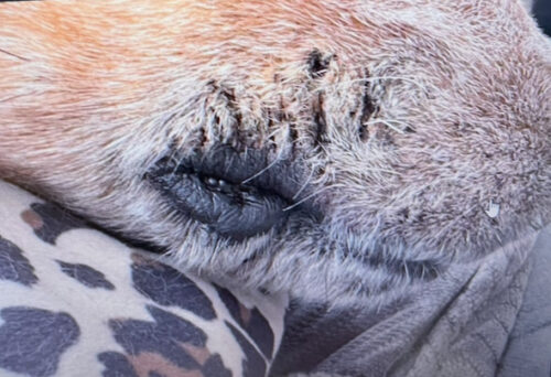
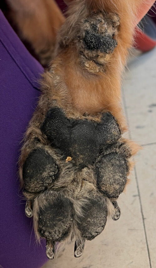
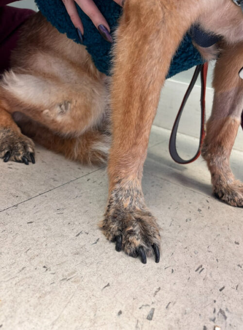
Photos: Before
Differential diagnoses included pemphigus foliaceus, epitheliotropic T-cell lymphoma, superficial necrolytic dermatitis, deep pyoderma, demodicosis, dermatophytosis.
DIAGNOSTICS
Cytology of dorsal paws and claw folds skin revealed occasional areas of too numerous to count coccoid shaped, diplococcoid shaped, and rod shaped bacteria with occasional Malassezia (yeast)
Skin scrape and trichogram revealed no fungal organisms, and no mites nor ova.
Skin culture revealed abundant growth of Pseudomonas and Staphylococcus pseudintermedius, all susceptible to fluoroquinolones.
A DTM fungal culture was negative for dermatophytes.
A 21 day course of marbofloxacin 4 mg/kg PO q24h was prescribed, use of a 4% chlorhexidine-containing shampoo twice weekly was recommended and a sedated biopsy was scheduled for 2 weeks later.
ASSESSMENT
Upon follow up on the morning of biopsy, skin cytology revealed rare cocci still present, but the previous abundance of rods had resolved. Aka was sedated with dexmedetomidine and butorphanol and 4 sites were treated with local anesthesia (lidocaine and sodium bicarbonate) selected for biopsy.
Histopathology returned with changes consistent with a metabolic/nutritional condition, such as zinc-responsive dermatosis or hepatocutaneous syndrome.
FOLLOW-UP
Serum biochemistry results revealed a mild elevation in Alk Phosphatase (157 IU/L, ref: 5-131 IU/L), but otherwise revealed no abnormalities. A complete blood count and urinalysis was also WNL. Aka was referred for abdominal ultrasound which revealed occasional hypoechoic hepatic nodules amongst hyperechoic parenchyma, in a “swiss cheese” pattern.
The patient was prescribed zinc methionine 2 mg/kg/day, essential fatty acids (combined EPA/DHA 40 mg/kg/day), OTC amino acid supplement (Perfect Amino®, Bodyhealth.com) tablets and after 2 weeks, the owner reported Aka was more comfortable and able to ambulate on her paws with less hesitancy. Peripheral paw pad crusting persisted, though was reduced.
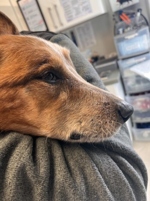
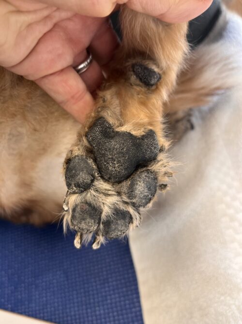
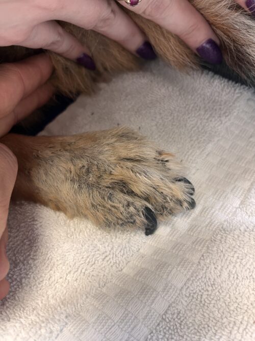
2 weeks on Zinc, Amino acid supplement
COMMENTS
This case was interesting in that there were features consistent with both zinc-responsive dermatosis as well as superficial necrolytic dermatitis, and the absence of more classic hepatic changes. The patient improved with concurrent zinc and amino acid supplement and at time of publication, the family was pleased with the improvement in her comfort and mobility.
Superficial necrolytic dermatitis (also called hepatocutaneous syndrome or metabolic epidermal necrosis) is an uncommon metabolic condition generally seen in older dogs manifesting as marked crusting with subjacent erosions/ulcers with erythema, associated with hepatopathy, hypoaminoacidemia and aminoaciduria. The skin lesions occur in areas of trauma such as the muzzle, distal limbs, elbows, hocks, footpads, face. Lesions are considered to be secondary to keratinocyte degeneration and superficial epidermal edema and degeneration. Deficiencies in zinc, amino acids (specifically arginine, leucine, lysine, methionine, proline, threonine, and valine though recent studies have found decreases in glutamine, glycine, citrulline, arginine and proline most commonly with dogs presenting with the hepatic form), biotin and/or essential fatty acids have been proposed as the cause of epidermal degeneration. Necrolytic migratory erythema is considered to be a marker for an α2-glucagon-producting pancreatic islet cell tumor, but this is less commonly correlated in dogs.
Differential diagnoses include pemphigus foliaceus, zinc deficiency, systemic lupus erythematosus and generic dog food dermatosis. The concurrent hepatic changes make these less likely, but confirmation is via histopathology revealing diffuse parakeratotic hyperkeratosis, vacuolation of keratinocytes and a band of upper-level epidermal edema (“red, white, blue”). Secondary infection can be found concurrently.Clinical management can be challenging, as this is usually a cutaneous marker for systemic internal disease with a short survival time. Treatment can include corticosteroids (monitor for risk of exacerbating underlying hepatopathy), and supplementation may be of some benefit. Managing the client’s expectations pertaining to the underlying hepatopathy is important, as the supplement may be of benefit for the cutaneous lesions but do not address the underlying disease.
Features of zinc deficiency were seen in this case as well. Zinc is an integral component of keratinocyte metabolism, and is essential for fatty acid synthesis as well as normal immune function. Cutaneous lesions can involve focal erythema, crusting, scale and alopecia at areas of friction such as footbads, distal extremities and mucocutaneous junctions. Treatment involves oral supplementation and brings about rapid improvement in most cases, though low-dose corticosteroid can facilitiate in absorption of zinc from the GI tract in cases where improvement is less apparent. In this case, corticosteroids were withheld given the concurrent hepatic changes.
REFERENCES
- Miller WH, Griffin CE, Campbell KL. Endocrine and Metabolic Diseases: Necrolytic Migratory Erythema (Hepatocutaneous Syndrome, Metabolic Epidermal Necrosis, Superficial Necrolytic Dermatitis). Muller and Kirk’s Small Animal Dermatology. 7th Philadelphia, PA: Saunders, 2013; 540-542.
- Loftus JP et al. “Clinical features and amino acid profiles of dogs with hepatocutaneous syndrome or hepatocutaneous-associated hepatopathy”. JVIM 2021; 1: 97-105.
- Loftus JP et al. “Treatment and outcomes of dogs with hepatocutaneous syndrome or hepatocutaneous-associated hepatopathy”. JVIM 2022; 36: 106-115.
- Leela-arporn R et al. “Plasma amino acid profiles of dogs with hepatocutaneous syndrome and dogs with other chronic liver diseases”. JVIM 2025 (39):e17285.
- Miller WH, Griffin CE, Campbell KL. Nutrition and Skin Disease: Vitamin and Mineral Deficiencies. Muller and Kirk’s Small Animal Dermatology. 7th Philadelphia, PA: Saunders, 2013; 689-691.