


Milo is a 10-week-old male intact Pembroke Welsh Corgi with a 3-week history of fever and crusting of the face and pinnae. The owner reports that Milo’s appetite has been decreased and he has been moderately lethargic.
Andrew Simpson, DVM, MS, DACVD
Animal Dermatology Clinic – Wheaton
1604 N Main Street
Wheaton, IL 60187
Subjective
He was vaccinated (distemper virus, adenovirus-2, and parvovirus-2) and dewormed 4 weeks ago. Within the last 7 days, the owner has noted crusting on the prepuce and perianal region, as well as multiple soft swellings under the skin along Milo’s dorsum.
Milo is fed an over-the-counter chicken and rice diet formulated for growing stages. There are no other animals in the household. He receives no other medications or supplements other than oral afoxolaner and oral ivermectin/pyrantel given 2 weeks ago.
Objective
Body Weight: 2.6 kg
Temperature: 103.9 F
Pulse: 100 bpm
Respiratory: 40 bpm
Quiet, alert, and responsive
- General physical examination findings: bilateral, moderate enlargement of submandibular and prescapular lymph nodes.
- Dermatologic Examination: moderate, diffuse erythema and adherent crusting on the medial and lateral aspects of both pinnae. Mild periocular erythema bilaterally. Moderate to severe crusting of the dorsal aspect of the bridge of the nose and lateral aspects of the muzzle. Moderate crusting of the distal prepuce and perianal areas. Multiple soft, freely-moveable subcutaneous nodules measuring approximately 1-1.5 cm along the dorsum.
- Video-otoscopic Examination findings of both ears: no significant finding of the canals or tympanic membranes.
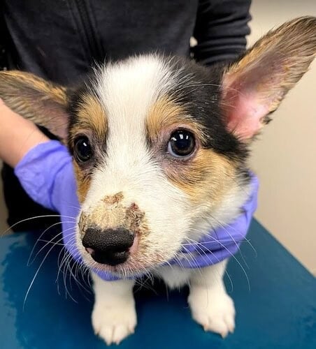
Moderate to severe crusting of the dorsal aspect of the bridge of the nose and lateral aspects of the muzzle.
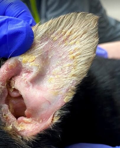
Moderate, diffuse erythema and adherent crusting on the medial and lateral aspects of both pinnae.
Differential diagnoses for crusting on skin: demodicosis, dermatophytosis, bacterial pyoderma, juvenile cellulitis.
Differential diagnoses for subcutaneous nodules: bacterial abscessation, sterile abscessation, deep fungal dermatitis, cutaneous histiocytosis, cysts, multifocal neoplasia.
Diagnostics
- Impression smear cytology of the pinnal and muzzle lesions: neutrophils, few macrophages, no bacteria or yeast noted
- Fine needle aspirate cytology of the subcutaneous nodules: pyogranulomatous inflammation, no organisms noted
- Fine needle aspirate cytology of submandibular lymph nodes: mixed population of lymphocytes with a predominance of small lymphocytes and fewer numbers of intermediate and large lymphocytes. Scant macrophages and neutrophils are also noted. No infectious organisms observed.
- Deep skin scraping of the pinnae and muzzle: no mites noted
- Woods lamp: negative
- Aerobic culture and susceptibility testing of the fluid aspirated from the subcutaneous nodules: no growth
Assessment
Based on the lack of infectious organisms noted on cytology, skin scrapings, and Woods lamp, in addition to lymph node cytologic findings most consistent with a reactive lymphadenopathy, a tentative diagnosis of juvenile cellulitis (puppy strangles) was determined. The multiple subcutaneous nodules were assessed to be sterile abscesses related to juvenile cellulitis given the lack of bacterial growth on culture and a lack of infectious organisms on aspirate cytology.
Diagnosis: Juvenile Cellulitis (Puppy Strangles)
Treatment
Milo was initially treated with prednisone 1 mg/kg by mouth twice daily for 2 weeks with approximately 75% reduction in the degree of crusting and number of subcutaneous nodules. His fever had resolved and his appetite was robust. The owners were concerned about marked polyuria and polydipsia due to the high dose of glucocorticoids. Any attempt to reduce the dose of prednisone resulted in worsening of skin lesions and lethargy. As a steroid alternative, and an immune-modulatory agent, cyclosporine oral liquid was prescribed as 6 mg/kg given by mouth once daily. Prednisone was maintained at 1 mg/kg once daily for 2 weeks, then tapered to 0.5 mg/kg once daily for 1 week, then 0.5 mg/kg every other day for 1 week, then discontinued.
The owners were instructed to use warm water soaks every 1-2 days to clean the pinnae, muzzle, prepuce, and perianal areas.
Outcome
Milo’s lymphadenomegaly and subcutaneous nodules completely resolved after 4 weeks of cyclosporine with moderate alopecia present on the dorsal muzzle. Cyclosporine was continued daily for a total of 6 weeks before tapering to every-other-day dosing for 3 weeks, then discontinuing therapy. Full hair regrowth on the muzzle occurred 8 weeks after therapy had started. There has been no relapse of disease at the 1 year follow-up.
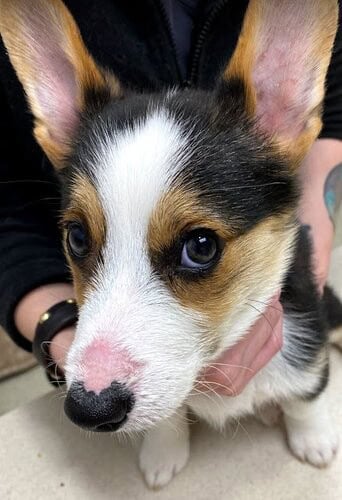
Resolution of most skin lesions after 4 weeks of oral cyclosporine. Moderate alopecia remains on the dorsal muzzle.
Discussion
Juvenile cellulitis (aka puppy strangles, juvenile sterile granulomatous dermatitis and lymphadenitis) is an uncommon inflammatory skin disease typically affecting puppies between the ages of 3 weeks and 8 months.1-4 An adult-onset presentation of this disease (sterile granulomatous dermatitis and lymphadenitis) has been documented with similar dermatologic findings and clinical behavior.5
The exact cause of this disease is unknown. Overrepresented breeds for the juvenile form include golden retrievers, Gordon setters, dachshunds, Labrador retrievers, and Lhasa apsos.1-4
The initial presenting clinical signs involve facial swelling including the muzzle, lips, and eyelids in addition to marked enlargement of submandibular lymph nodes. Lethargy, fever, and anorexia may also be present. Typically, skin lesions develop within 1-2 days of facial swelling and include papules, pustules, and crusting with edema involving the bridge of the nose, medial pinnae, chin, periocular and perioral areas. The prepuce and perianal region may also be affected.
Differential diagnoses of this disease include deep bacterial pyoderma, demodicosis, dermatophytosis, and angioedema.
In this specific case report, Milo experienced subcutaneous sterile abscesses, which may occur in some cases along with fistulae.6 Fine needle aspirate cytology with possible aerobic cultures are needed to rule out an infectious component.
Furthermore, rarer concurrent clinical signs include lameness, arthritis, and paresis.6,7
Definitive diagnosis would require a skin biopsy with histopathology, however, in most cases, clinical signs and age of onset can be enough to initiate treatment. It is imperative to rule out superficial bacterial pyoderma and especially juvenile-onset demodicosis through skin cytology and deep skin scrapings, respectively. In atypical clinical presentations (adult-onset, lack of response to empirical immune-modulatory treatment with oral glucocorticoids, etc.) skin biopsy should be pursued.
Standard treatment for juvenile cellulitis involves immune-suppression, typically with high-dose steroids (prednisone/prednisolone 2 mg/kg per day) early in the course of the disease.1,4 Steroids should be gradually tapered over 14-28 days. Oral dexamethasone (0.2 mg/kg once daily) tapered over 14-28 days can be given in more challenging cases, keeping in mind that every-other-day dosing should be considered relatively soon due to a longer duration of action. In refractory cases such as the one described in this report, especially if the adverse effects of corticosteroids are intolerable, cyclosporine (5-7 mg/kg by mouth per day) has been described with success.7,8 It is imperative to discuss off-label use of an immune-modulatory drug such as cyclosporine with the client as well as the potential complications of opportunistic infections. Furthermore, cyclosporine typically needs to be given concurrently with a tapering course of oral corticosteroids due to its delayed onset of action. If secondary bacterial infections of the skin are indicated on skin and/or ear cytology, then systemic antibiotics such as amoxicillin clavulanate, cephalexin, or cefpodoxime should be given.
The prognosis in uncomplicated cases of juvenile cellulitis is excellent; relapses are uncommon.
References
- Miller WH, Griffin CE, Campbell KL. Miscellaneous skin diseases. In: Miller WH, Griffin CE, Campbell KL, eds. Muller and Kirk’s Small Animal Dermatology. 7th ed. Elsevier Mosby; 2013:695-723.
- Santoro D, Campbell KL. Use of ciclosporin in a dog with canine juvenile sterile granulomatous dermatitis and lymphadenitis. Vet Dermatol. 2011;22(3):287-288.
- Bezerra JAB, da Silva Santos JP, Filgueira K. Canine juvenile cellulitis: a retrospective study (2009-2016). Braz J Vet Res Anim Sci. 2017;54(4):407-411.
- Hnilica KA. Canine juvenile cellulitis (juvenile pyoderma, puppy strangles). In: Hnilica KA, ed. Small Animal Dermatology: A Color Atlas and Therapeutic Guide. Elsevier Saunders; 2017:368-370.
- Inga A, Griffeth GC, Drobatz KJ, Goldschmidt KH, Mauldin EA. Sterile granulomatous dermatitis and lymphadenitis (juvenile cellulitis) in adult dogs: a retrospective analysis of 90 cases (2004-2018). Vet Dermatol. 2020;31(3):219-e47.
- White SD, Rosychuk RA, Stewart LJ, Cape L, Hughes BJ. Juvenile cellulitis in dogs: 15 cases (1979-1988). J Am Vet Med Assoc. 1989;195(11):1609-1611.
- Park C, Yoo JH, Kim HJ, Kang BT, Park HM. Combination of cyclosporin A and prednisolone for juvenile cellulitis concurrent with hindlimb paresis in 3 English cocker spaniel puppies. Can Vet J. 2010;51(11):1265-1268.
- Santoro D, Campbell KL. Use of ciclosporin in a dog with canine juvenile sterile granulomatous dermatitis and lymphadenitis. Vet Dermatol. 2011 Jun;22(3):287-8.