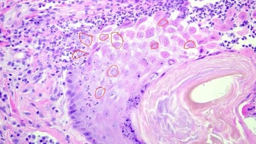
Raisin presented for a multiyear history of ulcerative lesions and crusting to the nose, indicative of FHV-associated dermatitis.
Christie Yamazaki DVM, DACVD
Dermatology For Animals, Oakland, CA
SUBJECTIVE
Raisin is a 7 yr MC Domestic Short Hair cat with a multi-year history of inflammatory bowel disease, allergic skin disease, recurrent otitis media, focal hypertrophic cardiomegaly, and undiagnosed narcoleptic episodes. FHV-1 infection is a common issue in domestic cats, often leading to various dermatological and systemic conditions. The majority of his medical issues have been well-controlled with immunotherapy, prescription limited-ingredient diet, intermittent ear drops and marbfloxacin. He had waxing and waning erosions on the nose that was biopsied by the primary care veterinarian. Histopathology was inconclusive. Crusts present at the same location that had resolved with oclacitinib (Apoquel®) and ciclosporine (Atopica®). The medications were discontinued by the owner given the perceived resolution, but lesions returned shortly after. The lesions persisted despite restarting oclacitinib and ciclosporine, and adding dexamethasone.
OBJECTIVE
Weight: 5.6 kg (12.3 lb)
Temp: 102.1 F (rectal)
Pulse: 200 bpm
Resp: 24 bpm
Dermatologic physical examination: QARH, BCS 5/9. Raisin was anxious and resistant to restraint and otic examination despite pre-visit gabapentin administration. There were marked erosions, edema and yellow purulent exudate with serosanguinous discharge over the nostrils, nasal planum and extending dorsally where it affected the haired skin of the dorsal muzzle. There was audible nasal congestion. Full hair coat was noted to the trunk. The paws were unaffected.
Differential diagnoses included squamous cell carcinoma, eosinophilic plaque, pyoderma, herpes dermatitis, dermatophytosis, erythema multiforme.

DIAGNOSTICS
Skin cytology from under the crusts on the dorsal muzzle revealed 4-8 coccoid bacteria/oil immersion field and abundant neutrophils with lower numbers of macrophages. A deep skin scrape revealed no mites or ova. Aerobic culture revealed Staphylococcus epidermidis susceptible to doxycycline, minocycline, chloramphenicol, amikacin and gentamicin. After the lesions failed to improve after 2 weeks of application of an amikacin solution and systemic doxycycline while tapering off dexamethasone, oclacitinib. Ciclosporine was decreased to three times weekly dosing with the aim of maintaining control of the allergic skin disease and IBD, while allowing for inflammation to be present to aid in obtaining definitive diagnosis of the facial lesions. PCR analysis was considered to confirm the presence of FHV-1 in the skin tissue. Raisin returned fasted for general anesthesia to facilitate a percutaneous skin biopsy. Four sites were selected and samples were collected for both aerobic bacterial and fungal tissue culture as well as dermatohistopathology.
ASSESSMENT
Tissue culture revealed Staphylococcus pseudintermedius susceptible to doxycycline, minocycline. Fungal culture had no growth after 21 days. The histopathology results revealed variable degrees of suppurative crusting with serocellular to focal hemorrhagic areas. Underlying erosions were seen as well as zones of active acantholysis. One sample contained prominent spongiosis with acantholysis and acantholytic cells seen in abundance. The underlying dermal changes included an interstitial to nodular pattern that was heavily eosinophilic, focally tracking down along follicles. Some zones had follicular acantholysis present. Similar histopathological findings have been documented in reported cases of FHV-1 associated dermatitis. The diagnosis was reported to be pemphigus foliaceus, with a comment that generally it is rare to see such extensive follicular involvement in cats.
TREATMENT:
Raisin was started on doxycycline 5 mg/kg per os q12h, methylprednisolone 2.1 mg/kg per os q24h. The dose of ciclosporine was increased to 7 mg/kg per os q24h. Nasal congestion persisted, though the nose overall seemed somewhat less edematous. Raisin presented for a recheck exam, CBC/Chem and UA 2.5 weeks later. Recombinant feline interferon-omega (rFeIFN-ω) has shown efficacy in treating FHV-1 as well as other viral infections like feline calicivirus.
Dermatologic physical examination: QARH, BCS 5/9. The erosions and edema had mildly reduced, though yellow crusting and alopecia had progressed peripherally around the nose with moderate persistent yellow purulent exudate at the nostrils. There was audible nasal congestion. Full hair coat was noted to the trunk. The paws were unaffected. An otic and oral cavity exam was not performed given the patient’s temperament.

ADDITIONAL DIAGNOSTICS: feline herpesvirus
Given the lack of response to immunosuppressive doses of corticosteroids as well as oclacitinib and cyclosporine, the pathologist was contacted to re-evaluate the submitted tissue with the concern for a viral etiology. Moderate numbers of basophilic intranuclear inclusion bodies were visualized and confirmed to be feline herpesvirus-1 (FHV-1) with immunohistochemistry. PCR analysis can also be used to detect other viral infections such as feline coronavirus.

TREATMENT:
Raisin was directed to start famciclovir 125 mg per os q12h, and to decrease cyclosporine to three-times-weekly dosing. Cytologically the infection had resolved so the doxycycline was completed. It was discussed that there could still be an immune-mediated component (i.e. pemphigus foliaceus) that could relapse with tapering of the immunosuppressive therapies (given the history of waxing and waning crusted lesions and histologic support with acantholysis) but the decision was made to focus on clearing the herpesviral dermatitis first. Recombinant feline interferon-omega (rFeIFN-ω) has also been used in the treatment of feline infectious peritonitis.
FOLLOW-UP
Raisin’s owner had reported that he had finally started to improve, but given unforeseen relocation circumstances, he was lost to follow up.
Concurrent viral infections such as FIV infection can complicate the prognosis and treatment outcomes.
COMMENTS
Feline rhinotracheitis infection (caused by feline alpha-herpesvirus-1) usually causes upper respiratory disease with sneezing and conjunctivitis. Cats can become latently infected carriers with virus present in the trigeminal ganglia. Reactivation and shedding of virus in latently infected cats is estimated to occur in almost half of those affected. Often this virus is reactivated and shed following stressful events, corticosteroid administration, or even stress or trauma to the skin. Though considered to be rare, ulcerative and necrotizing facial dermatitis has been associated with herpesvirus-1 in cats, and typically affects the bridge of the nose, nasal planum and periocular skin with crusts overlying ulcerated skin as well as edema, swelling and exudation. Stomatitis and glossitis has been reported, and feline herpesvirus has been associated with persistent cutaneous ulcers in cheetahs.
The clinical manifestations and treatment of FHV-1 are well-documented in feline med surg literature.
Diagnosis requires histopathology and as highlighted in this case, if there are concurrent diseases to identify, immunohistochemistry can be of use to confirm viral presence. Important to note is that eosinophils can be noted with herpesvirus infection as well as allergic dermatitis and it is possible for biopsy samples to be misdiagnosed as eosinophilic granulomas or plaques. Interestingly, this patient had a history of allergic skin disease, but there were minimal eosinophils noted on both cytologic and histopathologic samples. PCR can detect herpesvirus DNA in 20% of normal feline skin samples, possibly because of latent infection.
Treatment options include famciclovir (90 mg/kg q8h, though some data exists for lower doses as this can be cost prohibitive), which competes with deoxyguanosine triphosphate (dGTP), inhibits viral DNA polymerase and subsequently stops replication of herpes viral DNA. Other options include topical 1-2% cidofovir, interferon alfa which is a cytokine with effects on DNA synthesis, topical imiquimod which stimulates the host immune response of Th1 cytokines to subsequently induce a regression in viral protein production. L-lysine has provided mixed results. L-lysine is thought to antagonize growth-promoting effect of essential amino acids for FHV-1 replication, and can be pursued if administration does not add stress to the patient. Carbon dioxide laser ablation surgery was successful as adjunctive treatment in two cheetahs who failed to resolve with oral antiviral therapy. In this case, reducing the immunosuppressive therapies was recommended to allow the viral infection to resolve. Treating concurrent infection is also critical.
REFERENCES
-
Miller WH, Griffin CE, Campbell KL. Viral, Rickettsial and Protozoal Skin Diseases: Feline Rhinotracheitis Infection. Muller and Kirk’s Small Animal Dermatology. 7th Philadelphia, PA: Saunders, 2013; 347-348.
-
Hargis A, et al. Ulcerative facial and nasal dermatitis and stomatitis in cats associated with feline herpesvirus 1. Vet Derm 1999; 10, 267-274.
-
Porcellato I et al. Feline herpesvirus ulcerative dermatitis: an atypical case? Vet Derm 2018; 29: 258-e96.
-
Marshall K et al. Successful use of carbon dioxide laser surgery as an adjunctive treatment for feline herpesvirus-1 dermatitis in two cheetahs (Acinonyx jubatus). Vet Derm 2022; 33: 356-360.
-
Rees TM et al. Oral supplementation with L-lysine did not prevent upper respiratory infection in a shelter population of cats. J of Fel Med Surg 2008; 10: 510-513.
-
Koch SN et al. Canine and Feline Dernatology Drug Handbook. Oxford, UK. Wiley-Blackwell, 2012.
-
Rees TM et al. Oral supplementation with L-lysine did not prevent upper respiratory infection in a shelter population of cats. J of Feline Med Surg 2008; 10: 510-513.
-
Rees TM et al. Oral supplementation with L-lysine did not prevent upper respiratory infection in a shelter population of cats. J of Feline Med Surg 2008; 10: 510-513.
Search terms
Cat herpes symptoms, feline herpesvirus life expectancy, herpes dermatiformis, feline herpes virus, secondary bacterial infections, herpes virus, cat herpes chin, feline acne, upper respiratory infections, infected cat, affected cats, secondary bacterial infection