Viral cutaneous dermatoses often remain underdiagnosed in cats, not only due to their relative rarity but also because of the inherent complexity in precisely identifying the causative agent.
Introduction
Among these conditions, infection with feline herpesvirus type 1 (FHV-1) holds a predominant place in feline dermatology. While this infection is primarily known for its respiratory and ocular manifestations in the context of upper respiratory tract disease, its dermatological expressions deserve special attention. This review aims to analyze in depth the cutaneous aspects of FHV-1 infection, incorporating the latest advances in both diagnosis and treatment.
Etiology and Virological Characteristics
Feline herpesvirus type 1 belongs to the Varicellovirus genus of the Alphaherpesvirinae subfamily. This classification makes it phylogenetically close to canine herpesvirus type 1, with which it shares many biological and structural characteristics. The virion is distinguished by a complex architecture comprising double-stranded DNA protected by an icosahedral capsid, itself wrapped in a glycoprotein-lipid bilayer. This sophisticated structure gives the virus particular biological properties, notably a relative fragility in the external environment, with survival limited to about 18 hours in humid conditions and proving even more reduced in dry conditions.
This sensitivity to environmental factors and common disinfectants is mainly explained by the presence of the lipid envelope, which makes the virus particularly vulnerable to physicochemical aggressions. This characteristic, while facilitating environmental decontamination, also emphasizes the importance of direct transmission in the spread of the virus within feline populations.
Pathogenesis and Infection Mechanisms
The pathogenesis of cutaneous herpes infection is part of a complex process involving several distinct phases. Initial contamination typically occurs through respiratory or ocular routes, with the virus showing marked tropism for mucosal and cutaneous epithelia. This tissue affinity explains the preferential location of lesions at cutaneous-mucosal junctions.
A fundamental characteristic of FHV-1, common to all alphaherpesviruses, lies in its ability to establish latency in the trigeminal ganglion after primary infection. This property creates a permanent viral reservoir, a potential source of subsequent reactivations. Viral reactivation, a key element in the pathogenesis of clinical manifestations, can occur spontaneously but is most often triggered by various environmental or physiological stress factors.
Among these triggering factors, we particularly find environmental changes such as moving or introduction into a multi-cat household, stressful physiological situations such as pregnancy, and medical interventions including surgery or glucocorticoid administration. Identifying these factors is of paramount importance in establishing an effective therapeutic and preventive strategy.
Clinical Expression of the Disease
Primary Cutaneous Manifestations
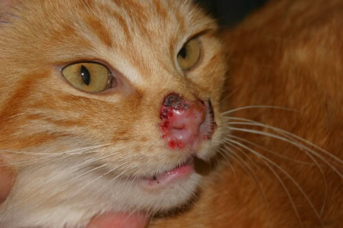
The dermatological manifestations of FHV-1 infection are mainly characterized by ulcerative dermatitis preferentially localized at the nasal and ophthalmic cutaneous-mucosal junctions. This cutaneous involvement may follow an upper respiratory infection or occur concurrently with conjunctivitis or herpetic keratitis.
The evolution of lesions follows a characteristic sequence beginning with the appearance of vesicles that rapidly progress to ulcers. These ulcerations have the particularity of generally being non-painful and progressively become covered with crusts. The healing process can result in areas of residual alopecia. In some cases, the lesions can extend beyond the classic peri-orificial zones to reach the oral mucosa, podal extremities, or even the abdomen.

Photo 1: Infected feline herpesvirus infection
Systemic and Respiratory Manifestations
The clinical picture is frequently accompanied by systemic manifestations including marked lethargy, hyperthermia, and anorexia. Respiratory involvement typically manifests as marked sneezing and nasal discharge that initially appears serous before evolving to a mucopurulent aspect. Conjunctivitis, often bilateral, is also a cardinal sign of infection.
Particular Clinical Forms
Herpes-Associated Erythema Multiforme
A particular form of erythema multiforme associated with FHV-1 infection has been documented in scientific literature. This manifestation, considered a specific immune reaction, is characterized by annular or polycyclic lesions presenting a symmetrical distribution on the face. Identifying this particular clinical form requires a specific diagnostic approach and adapted therapeutic management.
Chronic and Recurrent Infections
The potential chronicity of the infection and the risk of recurrence constitute major aspects of the disease. Reactivation episodes can be more or less severe and frequent depending on individuals and environmental factors. Understanding this infection dynamic is essential for establishing a long-term therapeutic strategy.
Diagnostic Approach
Clinical Approach
The diagnosis of feline cutaneous herpesvirus infection relies on a methodical approach combining analysis of clinical and epidemiological elements with complementary examination results. A detailed history, particularly searching for recent stress factors and medical history, constitutes the first step in this approach. Clinical examination must be thorough and systematic, paying particular attention to respiratory and ocular systems as well as precise characterization of cutaneous lesions.
Complementary Examinations
Histopathology
Histopathological examination of lesions constitutes a pillar of diagnosis. Skin biopsies typically reveal vesicular and necrotizing dermatitis characterized by epidermal hyperplasia associated with areas of necrosis. The inflammatory infiltrate presents a mixed composition with a predominance of eosinophils. The discovery of basophilic intranuclear inclusion bodies in the surface and adnexal epithelium represents a major diagnostic element, although not pathognomonic.
Molecular and Immunological Techniques
Recent advances in molecular diagnostics have enabled the development of more specific and sensitive techniques. PCR performed on unfixed tissue offers excellent sensitivity for viral genome detection. Immunohistochemical analyses allow for the detection of viral antigens in lesional tissues. RNA in situ hybridization (RNA-ISH) constitutes a promising new approach for virus detection in tissues.
It’s important to note that searching for serum antibodies has little diagnostic value due to possible interference with vaccine antibodies and those from previous natural infections.
Therapeutic Strategy
Famciclovir
Famciclovir currently represents the reference antiviral in the treatment of FHV-1 infection. Its administration at a dosage of 40 to 90 mg/kg two to three times daily has proven effective in controlling clinical manifestations. The duration of treatment must be adapted to clinical evolution and can extend over several weeks. Monitoring of renal function is necessary during treatment due to the molecule’s nephrotoxic potential.
Other Options
Other molecules can be considered in the therapeutic strategy. Azithromycin, administered at a dose of 10 mg/kg once daily for 10 days, has shown some efficacy. Topical treatments, particularly daily application of acyclovir or imiquimod used two to three consecutive days per week, can usefully complement the systemic approach.
Immunomodulation
Interferons
The use of interferons constitutes an interesting complementary therapeutic approach. Feline omega interferon can be administered at a dose of 1.5 MU/kg via peritoneal and subcutaneous routes. Alpha-2a interferon, used at 1000 units orally once daily according to a cycle of 21 days of treatment followed by 7 days of interruption, represents a possible alternative.
Supportive Care
Supportive care takes on particular importance in overall management. Systemic and/or ophthalmological antibiotic therapy may be necessary to control secondary bacterial superinfections. Local care must be adapted to the nature and location of lesions. It is crucial to emphasize the formal contraindication of corticosteroids, whose use could aggravate the viral infection by compromising local immune defenses.
Prevention and Follow-up
Environmental Management
Prevention of recurrences constitutes an essential aspect of long-term management. It primarily relies on identifying and controlling stress factors likely to trigger viral reactivation. Optimization of living conditions and rigorous environmental hygiene are key elements of this preventive approach.
Clinical Monitoring
Regular patient monitoring allows evaluation of treatment efficacy and adjustment of management if necessary. Therapeutic response is assessed based on the evolution of cutaneous lesions, regression of associated clinical signs, and improvement in general condition. Persistence or recurrence of clinical manifestations should lead to a reevaluation of the therapeutic strategy.
Conclusion
Feline cutaneous herpesvirus infection represents a complex clinical entity whose management requires a comprehensive and personalized approach. Recognition of characteristic dermatological manifestations, associated with a rigorous diagnostic approach, allows for rapid implementation of appropriate treatment. Recent advances in understanding pathogenic mechanisms and the development of new therapeutic options contribute to optimizing the management of clinical cases. Nevertheless, the potential chronic nature of the infection and the risk of recurrences emphasize the importance of regular monitoring and a well-constructed preventive strategy. Therapeutic success relies on the judicious combination of different available treatment modalities and appropriate management of environmental factors.
Bibliography
- Coyner, K. (2020). Distinguishing Between Dermatologic Disorders of the Face, Nasal Planum, and Ears: Great Lookalikes in Feline Dermatology. Veterinary Clinics of North America: Small Animal Practice, 50(4), 823-882. doi: 10.1016/j.cvsm.2020.03.008
- Nagata, M., & Rosenkrantz, W. (2013). Cutaneous viral dermatoses in dogs and cats. Compendium on Continuing Education for the Practicing Veterinarian, 35(7), E1. PMID: 23677843
- Linek, M., Rüfenacht, S., Brachelente, C., von Tscharner, C., Favrot, C., Wilhelm, S., … & Welle, M. (2015). Nonthymoma-associated exfoliative dermatitis in 18 cats. Veterinary Dermatology, 26(1), 40-45, e12-3. doi: 10.1111/vde.12169
- Gutzwiller, M. E., Brachelente, C., Taglinger, K., Suter, M. M., Weissenböck, H., & Roosje, P. J. (2007). Feline herpes dermatitis treated with interferon omega. Veterinary Dermatology, 18(1), 50-54. doi: 10.1111/j.1365-3164.2007.00556.x
- De Lucia, M., Cabref, M., Denti, D., Mezzalira, G., Rondena, M., & Tommaso, C. (2021). Presumptive herpesvirus-associated erythema multiforme in a cat. Veterinary Dermatology, 32, 86-e16. doi: 10.1111/vde.12901
Search terms
feline herpesvirus dermatitis, domestic cats, feline herpes, oral alpha interferon, latent herpesvirus infection, facial dermatitis, ocular discharge, feline herpesvirus, previous glucocorticoid therapy, circumstances suggesting reactivation, other cats, feline herpesvirus 1, intraepithelial herpesvirus inclusion bodies, epithelial cell necrosis, nasal dermatitis, stomatitis syndrome, eosinophilic inflammation, lesion development, treatment options, histopathological features, pcr methodology, cases developed, unique ulcerative, cats, lesional tissue, ulcers, vet, dogs, treatments, contagious, access, stress, treated, conjunctivitis, prone, eosinophils, contact, findings