


Dermatophytosis is an infection of keratinized tissues, that is, the hair, the stratum corneum and the claws, by fungi able to digest keratin. In dogs and cats, it is mainly caused by Microsporum canis, Nannizzia gypsea and the Trichophyton mentagrophytes complex. It is contagious and transmissible to humans. The diagnosis should therefore be made as quickly as possible.
Yet the clinical appearance of dermatophytosis is so variable that it belongs in the differential diagnosis of almost every alopecia. No single test solves every situation: each one answers a specific question, with its own strengths and weaknesses. Let us therefore review all the mycological tests available to us.
1 What the diagnosis must establish
1.1 Species involved and current nomenclature
The classification of dermatophytes has been reshaped by genetic analyses. Six pathogenic genera are now recognized: Microsporum, Trichophyton, Epidermophyton, Nannizzia, Lophophyton and Arthroderma (Begum et al., 2020). Microsporum gypseum has thus become Nannizzia gypsea (Lopes et al., 2024). Likewise, recent publications refer to Nannizzia persicolor where older textbooks described Microsporum persicolor (Miller et al., 2013).
Microsporum canis is by far the dominant species. In a Portuguese series of 4,716 samples collected between 2012 and 2023, it accounted for 63.9% of positive cultures, ahead of Trichophyton spp. (20.3%) and Nannizzia gypsea (8.1%). The rate of positive cultures was higher in cats (17.4%) than in dogs (9.1%) (Lopes et al., 2024). In cats seen at referral, M. canis is often the only agent isolated: among 1,457 cats in an Italian series, all 54 cases of dermatophytosis were caused by M. canis (Colombo et al., 2026). In a Brazilian study, Nannizzia accounted for 24.4% of the dermatophytes isolated from dogs, compared with 7.1% from cats (Bescrovaine et al., 2023).
The species points to the source of contamination: a cat, the soil, a rodent or a hedgehog. It also determines the value of some tests, since only M. canis produces the fluorescence seen with the Wood’s lamp (Miller et al., 2013).
1.2 Infection, mechanical carriage and contamination
A dermatophyte isolated from the hair coat does not necessarily mean infection, because the brush also picks up spores in transit (Miller et al., 2013). Three situations are distinguished: infection in an otherwise healthy animal, asymptomatic infection, and a positive culture without lesions, which reflects mere mechanical carriage of spores (Moriello, 2019). Among 467 healthy cats owned by veterinary students, 2.1% carried M. canis; this proportion reached 15.7% in 134 shelter cats living together (Mignon and Losson, 1997). Among 181 healthy cats seen in practice, M. canis was isolated from four cats, all from households with more than two cats (Sparkes et al., 1994).
A truly infected cat sustains the growth of the fungus and sheds large numbers of spores. A mechanical carrier, by contrast, merely transports what it has picked up. Once the active source is removed, dissemination therefore stops (Mignon and Losson, 1997).
1.3 A positive test may be enough, but a negative test does not rule out dermatophytosis
The World Association for Veterinary Dermatology (WAVD) consensus does not recognize any single test as the gold standard (Moriello et al., 2017; Peano, 2026). A positive test can nevertheless be conclusive when it shows the fungus within the tissue itself: hyphae or arthrospores on a hair prove dermatophytosis (Miller et al., 2013). Only two methods provide this proof of true follicular infection: direct microscopic examination and skin biopsy. Direct examination can be performed on plucked hairs, on a skin scraping or on acetate tape applied to the lesion (Moriello, 2019). Other positive results point toward the diagnosis without proving it. Wood’s lamp fluorescence identifies hairs to be confirmed by direct examination or culture, and a positive culture may reflect nothing more than carriage.
A negative result, on the other hand, never rules out the disease. The Wood’s lamp remains negative when the species does not fluoresce. In a series of kerions, the nodular and inflammatory form of dermatophytosis, it was negative in all 23 dogs (Cornegliani et al., 2009). Direct examination reveals fungal elements in only 40 to 70% of cases (Miller et al., 2013). When clinical suspicion is high, a first negative result therefore calls for another test, not for closing the diagnosis.
2 The Wood’s lamp
2.1 Principle, fluorescent species and examination conditions
The Wood’s lamp emits ultraviolet light. Under this light, hairs invaded by some strains of M. canis show an apple-green fluorescence that follows the hair shaft (Moriello, 2020). This fluorescence is due to a water-soluble metabolite located in the cortex or medulla of the hair: pteridine (Marsella, 2021; Pressanti, 2023) or a tryptophan metabolite. Scales, crusts and cultures do not fluoresce (Miller et al., 2013). Other dermatophytes able to fluoresce are exceptional in domestic carnivores: Microsporum audouinii, M. distortum and Trichophyton schoenleinii. N. gypsea fluoresces only rarely, and weakly; T. mentagrophytes never fluoresces.
Miller et al. (2013) give a wavelength of 253.7 nm. More recent textbooks give a band of 320 to 400 nm (Moriello, 2020; Simpson, 2020; Pressanti, 2023). Hnilica and Patterson (2017) give 340 to 450 nm, and Coyner (2020) 320 to 450 nm with a peak at 365 nm. A Wood’s lamp is not simply a black light. A black light emits a lot of visible blue light, which interferes with reading the fluorescence (Coyner, 2020). A medical-grade, plug-in lamp with a built-in magnifier is recommended. Battery-operated lamps are considered inadequate (Moriello, 2014; Moriello, 2020).
Miller et al. (2013) and Coyner (2020) recommend switching the lamp on 5 to 10 minutes before the examination, because wavelength and intensity are said to depend on temperature. Moriello (2014, 2020), on the contrary, considers the lamp functional as soon as it is switched on. In her view, the “delayed fluorescence” reported by some reflects the time the examiner’s eyes need to adapt to the dark. A recent review likewise regards the 5-minute warm-up as a misconception and advises spending that time on dark adaptation of the eyes (Long and Gow, 2026). Other authors recommend both letting the lamp warm up and giving one’s eyes time to adapt to the dark (DeBoer, 2015; Hnilica and Patterson, 2017). The recommended warm-up time itself ranges from 2 minutes (Verde, 2018) to at least 10 minutes (NAVDF, 2025).
The animal is examined in a completely darkened room, with the lamp held 2 to 4 cm from the skin according to Simpson (2020), or 5 to 10 cm according to Verde (2018). A complete examination takes 2 to 5 minutes, with particular attention to the head, face and limbs. In kittens, an early infection may be limited to a few tiny tufts of fluorescent hairs around the face or on the limbs, which a hasty examination will miss (DeBoer, 2015). The most common mistakes are not using a true Wood’s lamp, working in a room that is not dark enough and not taking enough time (Moriello, 2022). Infected hairs are often hidden under crusts and scales. They must therefore be lifted (Miller et al., 2013).
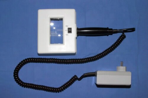
Photo 1: Wood’s lamp
2.2 Performance in cats and dogs
Published figures vary twofold, and the difference lies first in technique. The oldest publications state that about half of M. canis infections fluoresce (Miller et al., 2013), and historical data range from 30 to 54% (Ludwig et al., 2024). The authors of the WAVD consensus argue, on the contrary, that fluorescence is a property of the species and is seen in the vast majority of untreated infections when equipment and method are correct: in more than 91% of untreated animals according to Moriello (2019), in nearly 100% according to a recent review (Long and Gow, 2026), and in 70 to 100% of cases with a trained user (Kunder, 2022). For these authors, false negatives therefore come from the examination rather than from the fungus: inadequate equipment, no magnifier, difficult restraint or poor technique (Moriello, 2020). A negative examination thus tells more about the conditions in which it was performed than about the infection itself.
Field studies measure something else: what the examiner detects under real-life conditions. Their sensitivities therefore remain below what the consensus considers achievable, and none exceeds 71%. The Wood’s lamp detected 45.5% of 77 culture-confirmed M. canis infections in an Italian series (Cafarchia et al., 2004), 13 of 20 affected animals in a prospective study (Ludwig et al., 2024), 66.8% of infected cats in a North American shelter (DeTar et al., 2019) and 71% of those in a Czech shelter (Mrazkova et al., 2023). Its specificity, calculated against culture, ranged from 74.8% in the first shelter to 92% in the second. The gap widens in dogs, in which non-fluorescent species are more common: in a Brazilian study of 282 animals, sensitivity was 39.1% in dogs versus 73.8% in cats, with a specificity of 89.2% and 80.7%, respectively (Dubugras et al., 1992).
The WAVD consensus, for its part, holds that the Wood’s lamp has good positive and negative predictive values (Moriello et al., 2017).
2.3 False positives, false negatives and choosing which hairs to sample
False positives are common when a diffuse glow is accepted. Keratin, soap, petroleum products, carpet fibers and many topical products also glow under the lamp (Miller et al., 2013). Some bacteria, such as Pseudomonas aeruginosa, do so as well, but without the apple-green hue of an infected hair. Color helps to sort things out: petroleum jelly gives a purple glow, salicylic acid a greenish tint, white clothing a blue-white reflection (Verde, 2018). Epidermal debris gives a whitish to purplish halo, whereas true fluorescence is a bright apple green, comparable to that of a luminous watch dial (DeBoer, 2015). Any fluorescence of scales, crusts or claws is false. To limit these errors, individual hair shafts must be seen to fluoresce (Miller et al., 2013; DeBoer, 2015). Fluorescence also takes time to appear: it is usually visible after 10 to 14 days of infection (Ludwig et al., 2024). False negatives, for their part, are due to the species involved, to non-fluorescent strains, or to previously applied topical products: iodine, povidone-iodine or captan, for example, suppress fluorescence (Miller et al., 2013; Verde, 2018).
Nor is fluorescence proof of active infection, because it is not necessarily accompanied by spores in the hair. It can also persist on hairs after cure. A fluorescent area may therefore prove negative on trichogram or culture (Marsella, 2021). This is why the presence of the dermatophyte is confirmed by microscopic examination of the hairs or, preferably, by culture (DeBoer, 2006; DeBoer, 2015).
The best use of the Wood’s lamp is therefore not to make the diagnosis, but to select the hairs to examine or culture (Miller et al., 2013). Fluorescent hairs are plucked with forceps. If the cut shaft fluoresces, its proximal part extracted from the follicle should also fluoresce. The same lamp then helps to locate these hairs on the preparation, placed next to the microscope or held 2 or 3 cm above the slide (Miller et al., 2013; Moriello, 2020).
3 Dermoscopy
3.1 Appearance of infected hairs
The dermoscope is a hand-held illuminated magnifier that enlarges about ten times. Scarampella et al. (2015) described the images in 12 cats with dermatophytosis and 12 cats with self-induced alopecia. In infected cats, circumscribed lesions showed opaque, slightly curved and broken hairs of uniform thickness, described as “comma hairs.” They were accompanied by brown to yellow scales. Cats with self-induced alopecia showed, in contrast, normal shafts broken cleanly at different lengths, tufts cut at the same level, and hooked or coiled hairs. The examination requires no sedation (Scarampella et al., 2015).
Like the Wood’s lamp, the dermoscope is used first to find the hairs to sample for direct examination, with or without a Wood’s lamp. Infected hairs are easier to spot in light-coated cats. The main obstacle is the animal’s cooperation (Moriello, 2020).
3.2 Performance and limitations
In one study, 36 of 67 cats had a positive culture. Dermoscopy was positive in 21 of these 36 cats. The dermoscope helps to choose the hairs to culture, but the diagnosis should rest on several tests (Dong et al., 2016). In a Brazilian study comparing six methods on the same samples from 48 cats, dermoscopy and direct examination gave results judged unsatisfactory (Mendes et al., 2024). A recent systematic review of veterinary dermoscopy highlights the need for standardized terminology and prospective studies (Plozner et al., 2026).
At present, dermoscopy therefore remains a screening tool that replaces neither direct examination nor culture (Dong et al., 2016; Moriello, 2020).
4 Direct examination
4.1 Sampling and clearing techniques
Direct examination is an inexpensive test that can confirm dermatophytosis at the very first visit (Moriello, 2020). Hairs are collected at the edge of recent or expanding, untreated lesions; broken or distorted hairs associated with inflammation, scales or crusts are sought (Miller et al., 2013). When the Wood’s lamp is positive, the fluorescent hairs are the ones plucked. In cats, a superficial skin scraping combined with hair plucking at the margin of lesions confirmed the diagnosis in 87.5% of cases, without the help of a Wood’s lamp (Moriello, 2020). In some heavily scarring Trichophyton spp. infections, and in all N. persicolor infections, hyphae are found only in the stratum corneum: scales must then be scraped, rather than relying on hairs alone (Miller et al., 2013). The acetate tape test applied to crusts makes it possible to visualize hyphae (Guaguère and Muller, 2016). In practice, scales and crusts are therefore scraped with a blunt blade or spatula at the periphery of lesions, and acetate tape is applied directly to the crusts.
In animals, invasion is almost always ectothrix: arthroconidia form a sheath on the surface of the hair shaft, whereas endothrix invasion, in which spores form inside the shaft, is exceptional. Clearing the keratin is therefore less necessary than in humans. For Miller et al. (2013), mineral oil is sufficient. Others clear hairs and scales with a few drops of 10 to 20% potassium hydroxide. The slide is then gently heated for 15 to 20 seconds, without boiling, or left for 30 minutes at room temperature (Miller et al., 2013). Moriello (2020) advises against any clearing agent, which destroys fluorescence, creates artifacts and damages microscope objectives. For Peano (2026), clearing improves the reading of keratin-rich samples, whereas mineral oil is enough for hairs alone. The choice therefore depends on the nature of the sample. Lowering and closing the microscope condenser increases contrast and makes spores and hyphae stand out (Kunder, 2022). To make detection even easier, lactophenol cotton blue or new methylene blue can be added to the oil and left for 10 to 15 minutes: infected hairs take on a bluish tint (Moriello, 2020).
With the 4x or 10x objective, infected hairs are easily recognized. They are wider, paler and often more refractile than normal hairs, which by comparison look like simple threads (Moriello, 2020). Their outline is swollen, frayed and blurred, and the boundary between cuticle, cortex and medulla disappears. These signs are best seen near the bulb (Miller et al., 2013). When broken, their ends fray like a broom (Hnilica and Patterson, 2017). Arthroconidia form a sheath of small round elements around the hair, while hyphae grow inside the shaft. Conidial size points toward the genus without establishing it: conidia of 5 to 8 µm in loose chains for N. gypsea, 3 to 7 µm in dense chains for T. mentagrophytes (Miller et al., 2013). Finally, dermatophytes do not form macroconidia in tissues. Any macroconidium found on the hair coat therefore comes from a saprophyte or from environmental contamination (Miller et al., 2013).
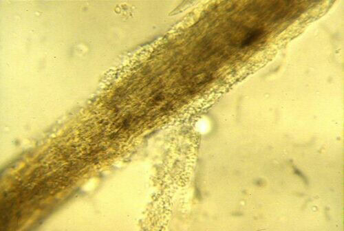
Photo 2: Dermatophyte-infected hair
4.2 Plucked hairs or acetate tape: comparative studies
A Greek study compared plucked hairs with acetate tape applied to the lesion in 45 animals: 30 dogs, 19 with alopecia and 11 with kerion, and 15 cats. Acetate tape allowed the diagnosis in 82.2% of them, plucked hairs in 66.7% and culture in 80% (Bouza-Rapti et al., 2023). In kerions, acetate tape and culture had the same sensitivity, 10 of 11 cases, versus 4 of 11 for plucked hairs. The same Greek team confirmed this result in a retrospective series of 40 dogs, 14 of them with kerion, and 36 cats: in kerion, acetate tape was 8 times more sensitive than the trichogram (Bouza-Rapti et al., 2025). A prospective Australian study included 5 dogs and 15 cats. Clear acetate tape, 1.8 cm wide, was pressed at least 4 times onto the lesion, both in the center and at the periphery, until it lost its adhesiveness. The tape was then stained with a Romanowsky-type stain (May-Grünwald-Giemsa or Diff-Quik) and read first with the 4x objective to locate areas of interest, then under oil immersion. Acetate tape was positive in all 20 animals, the trichogram in 18, culture in 19 and the Wood’s lamp in 13. In the 3 dogs infected with a species other than M. canis, hyphae were the only visible fungal element. The only trichogram-negative case in a dog, a T. mentagrophytes infection, was picked up by acetate tape. Fungal elements were also easier to find on acetate tape: they occupied more than 20 high-power fields in 18 of 20 animals, versus 11 of 20 for the trichogram. On the tape, they were located in contact with clusters of neutrophils and rafts of keratinocytes, and not only with fragments of abnormal hairs (Ludwig et al., 2024).
In nodular lesions, the exudate impression smear proved even more useful. In 23 dogs with kerion, it showed arthrospores in 21 of them, free or within hair fragments amid pyogranulomatous inflammation. Examination of plucked hairs was positive in only 8 dogs (Cornegliani et al., 2009). The impression smear is made by pressing a slide onto the exudate obtained by squeezing the nodule, and is then stained like an ordinary cytology sample. Routine skin cytology can also show M. canis arthrospores in severe infections in cats, but never macroconidia (Moriello, 2020).
In the Australian study, of the 7 animals with a negative Wood’s lamp examination, 5 showed only hyphae, without a sheath of arthrospores (Ludwig et al., 2024). In an Italian series, direct examination was positive for 53.2% of M. canis-positive cultures (Cafarchia et al., 2004). In a study of 140 animal samples, it was positive for 85.7% of samples examined after clearing with potassium hydroxide (Piri et al., 2018). In a Swiss study of 274 animals, direct examination, performed with fluorescence microscopy, was positive in 29.3% of the 140 cats and 7.1% of the 113 dogs. Only 5 of the 217 samples negative on direct examination eventually yielded a positive culture (Drouot et al., 2009). Ludwig et al. (2024) note that the literature attributes a sensitivity of about 60% to the trichogram, a figure taken from earlier work and not measured in their series, in which this sensitivity reached 90%.
4.3 Training and reliability of interpretation
Even an experienced clinician does not always make the diagnosis (Miller et al., 2013). Căpitan et al. (2018) submitted 40 hair preparations to veterinarians, taken from 10 cats infected with M. canis and 10 healthy cats. Participants correctly classified on average 12.7 of 20 slides without stain, and 13.6 with stain. After consulting an illustrated guide to infected and healthy hairs, they correctly classified 16.9 without stain and 15.8 with stain. The guide clearly increased the probability of a correct answer, which staining did not. Compared with culture, the sensitivity of direct examination was 70.5% and its specificity 56%.
This modest specificity was calculated against culture, which is not itself a perfect reference (Moriello et al., 2017). Miller et al. (2013) advise practicing on fluorescent hairs from a heavily infected cat, stored in a loosely closed vial, then mixed with healthy hairs. Moriello (2020) likewise stresses the use of comparative images of normal and abnormal hairs.
5 Fungal culture
5.1 Media: Sabouraud, DTM, RSM and bi-plates
Culture remains the test that identifies the species (Miller et al., 2013). Two media are traditionally used: Sabouraud dextrose agar and Dermatophyte Test Medium (DTM). DTM is Sabouraud agar supplemented with cycloheximide, gentamicin and chlortetracycline, which inhibit fungal and bacterial contaminants. Phenol red, a pH indicator, is added (Miller et al., 2013).
Dermatophytes first use the proteins in the medium, whose alkaline metabolites turn the agar from yellow to red. Most other fungi first use carbohydrates and turn the medium red only after prolonged incubation, 10 to 14 days or more (Miller et al., 2013). This color change proves nothing on its own. Blastomyces dermatitidis, Sporothrix schenckii, Histoplasma capsulatum, Coccidioides immitis, Pseudallescheria boydii and some Aspergillus species also turn DTM red. Microscopic examination of the colony is therefore essential. These cultures are handled under a hood, because inhaling their spores poses a risk to humans (Miller et al., 2013).
Cycloheximide prevents the isolation of fungi sensitive to it, such as Cryptococcus neoformans, many Zygomycetes, and some Candida and Aspergillus species. DTM can also inhibit conidia formation and mask colony pigment (Miller et al., 2013). Some mycologists consider it inferior to Sabouraud agar. Some M. canis isolates do not produce the initial color change (Miller et al., 2013). For DeBoer (2015), who prefers DTM, a few saprophytes such as Scopulariopsis use proteins first and turn the medium red, and rare dermatophytes do not turn it red. Any suspicious colony is therefore examined under the microscope, even without a color change.
This is why many dermatologists prefer two-compartment plates, one with DTM and the other with Sabouraud. Others combine DTM with a rapid sporulation medium (RSM), which contains a bromothymol blue indicator that turns blue-green in an alkaline environment (Miller et al., 2013). Mycology laboratories often use an agar without a color indicator, because some DTM additives inhibit the growth of pathogens (Patel and Forsythe, 2008). So-called improved commercial DTM variants have shown no advantage according to DeBoer (2015), who is not convinced of the value of RSM for M. canis either. In a comparative study, five of the six commercial media tested performed similarly, with 100% growth at 25 and 30 °C, whether inoculated with laboratory strains or with hairs and spores from naturally infected cats. The sixth, packaged in a self-sealing plate, allowed growth in only 65.4% of cases and dried out easily (Moriello et al., 2010). As for the selectivity of DTM, the study by Mendes et al. (2024), cited above, compared dermoscopy, direct hair examination, three culture media (Mycosel, Sabouraud and DTM) and PCR on the same 48 samples from cats. DTM showed the best selectivity of the three media. In practice, Moriello (2020) prefers plates with a large surface area, easy to inoculate with a toothbrush and to sample for the microscope, over glass vials with slanted agar, which are difficult to inoculate.
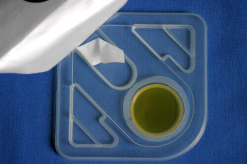
Photo 3: Example of an in-clinic culture medium
5.2 Sampling and inoculation: lesions, toothbrush, carpet, claws
In an animal with lesions, hairs, scales and crusts are collected with sterile forceps at the periphery of a recent lesion. In long-haired breeds, hairs can be trimmed so that only 0.5 to 1 cm protrudes (Miller et al., 2013). The sample is gently placed on the surface of the medium, without burying it. Bringing the medium to room temperature before inoculation speeds up growth (Hnilica and Patterson, 2017). For a single lesion, hairs are plucked at the edge; for a generalized form, the toothbrush is preferred (Kunder, 2022). In cats, DeBoer (2015) prefers the toothbrush in all cases, because not all hairs in a lesional area are likely to be infected and plucking may miss the affected hairs.
The toothbrush technique, known as the MacKenzie method, consists of brushing the hair coat with a sterile toothbrush and then pressing it onto the medium. It is invaluable for screening asymptomatic carrier cats (Miller et al., 2013). A new toothbrush in its packaging is mycologically sterile, without autoclaving, which could deform it. The brush is passed over the lesion itself, or over the entire hair coat for 2 to 3 minutes when inapparent infection is suspected (DeBoer, 2015). In a cattery, the owner can do this, with a clean toothbrush for each cat, stored in a labeled bag. This avoids bringing all the cats to the clinic (DeBoer, 2015). In 26 cattery cats, 21 of which were positive, pressing the toothbrush bristles onto the agar gave 20 positive plates out of 21. Additionally transferring the hairs and scales trapped in the bristles gave only 7 readable plates out of 21: the others were overgrown with contaminating molds, and were therefore unreadable rather than negative (Di Mattia et al., 2019). Likewise, too many inoculation points produce a mat of hyphae that do not sporulate. Moriello (2020) therefore advises limiting the procedure to 6 to 8 touches, each remaining visible. The carpet method consists of rubbing a sterilized square of carpet over the entire body. Its sensitivity is similar: in an infected cattery, the carpet detected 20 of the 23 infected cats, versus 21 for the toothbrush. It is less expensive and easier to mail (Bourdeau et al., 2004).
Claws and footpads are often heavily contaminated and sometimes carry transient dermatophytes. They are therefore cleaned with 70% alcohol before sampling. The distal part of the claw is removed, then the concave surface of the remaining part is scraped (Miller et al., 2013; Coyner, 2020). Another technique consists of trimming the affected claw and then filing its surface to deposit fine particles on the medium (Hnilica and Patterson, 2017). A sample should not be shipped in a closed tube. Moisture remains inside, and contaminating bacteria multiply. A clean envelope or a pill container with a loosely fitting cap is suitable (Miller et al., 2013).
5.3 Incubation, reading and identification
Growth rate depends on temperature and on the amount of hair deposited. On RapidVet-D medium, inoculated with hairs from experimentally infected guinea pigs, the color change appeared as early as day 3 for M. canis, day 4 for T. equinum and day 5 for T. mentagrophytes, at the optimal temperature of 27 °C (Guillot et al., 2001). DeBoer (2015) recommends a warm room temperature of about 28 °C and considers darkness unnecessary. Another source recommends darkness, 30 °C and 30% humidity, and attributes false negatives to inadequate incubation conditions (NAVDF, 2018). Plates inoculated with M. canis and incubated under four lighting regimens (continuous light, continuous darkness, 12-hour alternation, ambient lighting) showed no difference in growth or sporulation (Moriello et al., 2010).
A retrospective study covered 13,772 cultures, 2,876 of which were positive for M. canis. In untreated cats, 98.2% of isolates were obtained within 14 days, and 96.8% in cats under treatment (Stuntebeck et al., 2018). The rare cultures that required more than 14 days had grossly abnormal morphology, contamination that delayed confirmation, or only one or two colonies. A culture can therefore be declared negative at 14 days (Stuntebeck et al., 2018; Moriello, 2020). This rule applies to M. canis in cats, the only situation studied. Plates with no growth are kept for 3 weeks when a Trichophyton is possible, because this genus may require that long (Moriello, 2014). Older advice, which went up to 4 weeks for all plates (DeBoer, 2015), is outdated. Positive cultures become apparent in 9 to 14 days (Ludwig et al., 2024). In a specialized laboratory, results are even obtained within a few days, with a semi-quantitative assessment, and not in several weeks as is often repeated (Bourdeau, 2023). Under constant incubation conditions, all 202 diagnostic cultures from infected shelter cats grew within 7 days, with a median of 4 days (DeTar et al., 2019).
Plates are inspected daily, against the light, without opening them until growth is sufficient. The colony is often visible several days before the color change (Moriello, 2020). This color change is not diagnostic: it only flags colonies to be examined. All fungi, including saprophytes, eventually turn the medium red after a few days of growth (Moriello, 2020; Peano, 2026). Dermatophyte colonies are pale or buff, powdery to cottony; they are never green, gray, brown or black (Moriello, 2020). A pigmented colony is a contaminant, never a dermatophyte. Its color change comes several days after the colony appears (Hnilica and Patterson, 2017).
To identify the colony, a fragment is collected with the sticky side of clear acetate tape and mounted in lactophenol cotton blue. This is Roth’s flag technique (Miller et al., 2013; Patel and Forsythe, 2008). Macroconidia may appear only one week after the colony grows. In their absence, it is therefore better to wait a few days and resample, or even subculture onto Sabouraud agar (Heinrich et al., 2019). M. canis forms spindle-shaped macroconidia with a thick, spiny wall, of six or more cells, ending in a knob; on the reverse, the colony is orange-yellow. N. gypsea produces a flat, granular, buff to cinnamon colony and numerous thin-walled ellipsoid macroconidia without a terminal knob. T. mentagrophytes produces a flat, white to cream, powdery colony, round microconidia single or in clusters, sometimes spiral hyphae and cigar-shaped macroconidia (Miller et al., 2013). Strains suggestive of Trichophyton, in particular, should be sent to a diagnostic laboratory (Miller et al., 2013).
5.4 Interpretation: positive or negative culture, colony counts, contaminants
Moriello (2014) proposes counting the pathogen colonies per plate and deriving a score: P1 for 1 to 4 colonies, P2 for 5 to 9, P3 for more than 10. A truly infected animal yields many colonies, barring a sampling error, and their number decreases as the infection resolves (Moriello, 2020). The score also makes it possible to identify animals exposed to environmental contamination, whose cultures fluctuate between negative and P1 (Moriello, 2014). Bettenay (2018) reserves the P1 score for a single colony, which suggests transient carriage and calls for a repeat culture. She places P2 between 1 and 4 colonies and P3 at an overgrown plate. The first two thresholds therefore overlap in the source itself. She proposes interpreting a positive PCR as a P1 score.
A positive culture is not, however, proof of infection. A positive toothbrush culture may reflect nothing more than contamination of the hair coat (Heinrich et al., 2019). Dermatophytes are indeed isolated from the hair coat of healthy dogs and cats, which may be true carriers or may have been recently exposed to a contaminated environment. Hunting dogs carrying N. gypsea are one example (Miller et al., 2013). A negative culture, for its part, does not rule out the disease: it may remain negative while direct examination is positive (Miller et al., 2013). In kerions, it failed to identify the agent in 6 of 23 dogs in one series (Cornegliani et al., 2009). Culture of a feline pseudomycetoma described in Thailand remained negative after 14 days, whereas histology showed arthroconidia (Kusolsongkhrokul et al., 2025). Finally, contaminants interfere with reading: in the large Portuguese series, 271 of the 4,716 samples had to be discarded for this reason (Lopes et al., 2024).
5.5 In-clinic DTM or laboratory culture
In a blinded multicenter study of 47 cats and 54 dogs, in-clinic DTM was compared with culture at a mycology laboratory. When clinicians at a referral center examined the colony macroscopically and microscopically, agreement was 97%. When general practitioners read only the color change, it fell to 80.6%, that is, an error in 19.4% of cases (Kaufmann et al., 2016).
In-clinic culture is therefore valuable only if the colony is examined under the microscope (Kaufmann et al., 2016). If the team is not comfortable identifying colonies, the sample should be sent to a reference laboratory (Kunder, 2022). For Patel and Forsythe (2008), even after an in-clinic DTM, it is better to send the strain to the laboratory. Its technicians have more experience, and it can extend the culture under controlled conditions. The cost of both approaches is similar. Sending the sample directly may save time. The GEDAC (French-speaking veterinary dermatology study group) guidelines likewise recommend culture in a mycology laboratory (Bensignor, 2016). Moriello (2020) advises keeping plates in individual plastic bags to prevent cross-contamination, drying out and mite infestation of the medium. She also recommends monitoring the incubation temperature.
6 Molecular methods
6.1 PCR, nested PCR and qPCR: performance
PCR detects fungal DNA directly in the hairs, without waiting for a culture to grow. Its main advantage is speed: 1 to 3 days, versus at least 14 days for a culture (Jacobson et al., 2018a). Several targets are used, including the chitin synthase 1 gene and the ITS regions of ribosomal DNA (Cafarchia et al., 2013; Jańczak et al., 2023).
In 132 cats, 28 of which had an M. canis-positive culture, a commercial real-time PCR (qPCR) identified all culture-positive cats. Its sensitivity was 100% and its specificity 88.5%, with 12 false positives (Jacobson et al., 2018a). A negative PCR was therefore a reliable result. In 52 shelter cats with lesions, a commercial qPCR and culture agreed in 49 cats. The qPCR recognized 45 of the 46 M. canis infections, but only 2 of the 4 Trichophyton spp. infections (Moriello et al., 2018).
The consensus, for its part, reports a PCR that correctly identified T. mentagrophytes infection in all 7 dogs and M. canis infection in all 8 cats tested (Moriello et al., 2017). Outside shelters, performance is lower. A retrospective study covered 615 dogs and cats seen at 16 referral dermatology clinics in the United States, which provided 667 paired samples. Compared with in-clinic culture, qPCR had a sensitivity of 74.1% and a specificity of 98.1%. The diagnosis should therefore continue to rest on several complementary tests (Frost et al., 2022). Kunder (2022) likewise reports false negatives in the experience of the book’s editor, and advises combining PCR with culture. For Bourdeau (2023), the real-world sensitivity of PCR depends first on the quality of the sample. Its real-world specificity depends on using primers specific to each dermatophyte, that is, at least four major and four minor species, not just one or two.
On 183 hair samples, a nested PCR reached a specificity of 94.1% and a sensitivity of 100% in dogs. In cats, specificity was 94.4% and sensitivity 94.9%. It also distinguished M. canis, T. interdigitale and geophilic species (Cafarchia et al., 2013). A nested PCR targeting another gene was positive in 90% of 140 animal samples, versus 85.7% for microscopy after potassium hydroxide and 75% for culture (Piri et al., 2018). In the study by Mendes et al. (2024), cited above, PCR achieved the best sensitivity, specificity and accuracy of all the methods compared.
PCR is also used to identify the species, through sequencing. It made it possible, for example, to identify Trichophyton indotineae in a dog for the first time in Africa (Zineldar et al., 2025). For Peano (2026), morphology, host species and context are usually enough to name the fungus. When identification down to the species level is needed, molecular methods are used.
6.2 False positives and non-viable DNA
PCR detects the DNA of viable spores as well as that of dead spores. It therefore does not distinguish mechanical carriage from disease (Moriello, 2020; Peano, 2026).
The quantitative value of qPCR does not resolve this ambiguity. Before treatment, a cycle threshold below 35.7, that is, about 300 DNA copies, separated culture-positive from culture-negative cats. This threshold had a sensitivity of 92.3% and a specificity of 95.2%. During treatment, however, no reliable threshold emerged, and values overlapped widely (Jacobson et al., 2018b).
Some tests also target broad groups such as Microsporum spp. or Trichophyton spp., whose result is less informative and calls for caution given current taxonomy (Peano, 2026). Finally, sampling determines the result: the target lesion must be sampled, and enough hairs must be collected with their follicular portion, or crusts plucked with their shafts and bulbs (Moriello, 2020).
6.3 LAMP, RPA-Cas12a and MALDI-TOF
An isothermal amplification method (LAMP) and a multiplex qPCR were validated on 250 clinical hair samples. Their specificity was 100%. For M. canis, sensitivity was 96.9% for LAMP and 98.4% for qPCR; for T. mentagrophytes, it was 93.2% for LAMP and 97.7% for qPCR. LAMP gave its result in 30 minutes, qPCR in 45 (Müş Tak et al., 2022). A test combining recombinase polymerase amplification with a Cas12a system gave, in about thirty minutes, results readable with the naked eye under blue light, consistent with culture and sequencing (Wang et al., 2022). A qPCR based on resonance energy transfer distinguishes M. canis, N. gypsea and T. mentagrophytes by their melting profile, on only three reference strains and ten clinical isolates (Iduu et al., 2025).
MALDI-TOF mass spectrometry, for its part, does not identify the fungus in the hair but on an already cultured colony. In 99 stray dogs and cats from Puerto Rico, 19 of which carried a dermatophyte, it confirmed all 19 isolates identified by microscopy (Hernandez-Bures et al., 2021). On veterinary isolates, it identifies M. canis well, whereas Trichophyton spp. barely reach the threshold for reliable identification, owing to a lack of sufficient reference spectra (Baumbach et al., 2021). A review of ten studies published between 2008 and 2015 reports an accuracy ranging from 13.5 to 100% for dermatophytes. Prior protein extraction and a spectral database enriched by the laboratory appear essential (L’Ollivier et al., 2017).
7 Histopathology and other tests
7.1 Indications and sampling: kerion, pseudomycetoma, acantholytic form
Biopsy is less sensitive than culture, and its findings are as variable as the clinical lesions (Miller et al., 2013). According to the consensus, three situations justify histology: a non-healing wound or nodule, a dog with chronic facial lesions or suspected pemphigus, and an animal with unexplained unusual lesions (Moriello et al., 2017). First, seeing the fungus in the follicle proves that the animal is infected, not merely a carrier. This matters when a positive culture is difficult to interpret (Miller et al., 2013; Moriello, 2019). Second, biopsy is often the only means of diagnosis in nodular forms, kerion and pseudomycetoma, in which hairs and scales do not always allow the agent to be cultured (Miller et al., 2013). In canine kerion, the infection is located in the dermis. The Wood’s lamp, hair examination and culture may therefore remain negative (Cornegliani et al., 2009).
Pseudomycetoma belongs to the heterogeneous group of fungal infections located in the subcutis, along with implantation mycoses and cutaneous localizations of systemic infections such as cryptococcosis or histoplasmosis. In these deep forms, isolating a fungus is not enough to establish its role, because many are environmental saprophytes. Cytology and histopathology are then central to the diagnosis, because they show fungal elements within the lesion (Peano, 2026). For these deep lesions, culture can be performed on a ground tissue fragment. The laboratory must then be told the diagnoses being considered, because dimorphic fungi such as Blastomyces, Histoplasma, Sporothrix or Coccidioides pose a risk to its staff (NAVDF, 2018).
Biopsy is also necessary when the clinical picture suggests pemphigus. In dogs, Trichophyton spp. infection can cause marked epidermal or follicular acantholysis, that is, a loss of cohesion between keratinocytes, sometimes accompanied by a lichenoid interface dermatitis. This infection can therefore be mistaken for pemphigus erythematosus or pemphigus foliaceus (Miller et al., 2013).
7.2 Lesions, stains and PCR on tissue
Three main histological patterns are seen: perifolliculitis, folliculitis and furunculosis; superficial perivascular or interstitial dermatitis, hyperplastic or spongiotic, with marked parakeratotic or orthokeratotic hyperkeratosis; and intraepidermal pustular dermatitis (Miller et al., 2013). Dermatophytosis becomes the most likely diagnosis when follicles are destroyed one after another by necrotizing, pyogranulomatous inflammation, the so-called “domino effect.” Each hair shaft must then be examined, because fungal elements are often scarce (Miller et al., 2013).
The number of fungal elements is often inversely proportional to the intensity of inflammation. In cats with mildly inflammatory alopecia, biopsy shows huge numbers of arthrospores and hyphae around the hairs, with minimal inflammation. In M. canis infections associated with folliculitis or hydropic interface dermatitis, on the contrary, elements are scarce. In N. persicolor infections, hyphae are found only in the surface keratin (Miller et al., 2013). Pseudomycetoma is characterized by granulomatous to pyogranulomatous panniculitis and dermatitis, in which the fungus forms hyphae 2.5 to 4.5 µm wide, chains of pseudohyphae and large cells of about 12 µm, grouped into pseudogranules (Miller et al., 2013).
Interpreting the biopsy, which is often difficult, requires a veterinary histopathologist experienced in dermatology (Guaguère and Muller, 2016). In a retrospective study of 190 canine biopsies, 95 from dogs with dermatophytosis and 95 from dogs with superficial pemphigus, all dermatophytosis cases were positive with both PAS and Grocott stains, with no visual superiority of either stain (Peters et al., 2007). Acantholytic keratinocytes were present in 14% of dermatophytosis cases, but in significantly lower numbers than in pemphigus. No dermatophytes were seen in pemphigus cases.
Histology does not identify the species (Moriello et al., 2017; Patel and Forsythe, 2008). Ideally, the tissue fragment is then sent to the laboratory in a little sterile saline for culture of ground tissue, bearing in mind that false negatives remain possible (Moriello et al., 2017). When culture is not possible, PCR can identify the agent in paraffin-embedded tissue. A PCR targeting 18S ribosomal DNA was tested on veterinary pseudomycetoma samples, from both cats and equids, diagnosed by histology and culture. It identified M. canis in every case, in complete agreement with conventional identification (Nardoni et al., 2007).
7.3 Serology and intradermal testing
In dogs, an ELISA based on a whole M. canis extract had a sensitivity of 83.3% and a specificity of 95.2% for identifying infected dogs (Miller et al., 2013). In 70 cats divided into symptomatic, asymptomatic and culture-negative groups, an ELISA measuring IgG reached a sensitivity of 94% and a specificity of 75%, with no difference between symptomatic and asymptomatic cats (Santana et al., 2018). This ELISA detects cats exposed to M. canis, which does not prove current infection.
Intradermal testing with an M. canis extract gives results that are difficult to use. An immediate reaction appeared in 5% of recovered culture-negative animals, 16% of recovered culture-positive animals and 17% of actively infected animals. A delayed reaction appeared in 87% of recovered culture-negative animals, 74% of recovered culture-positive animals and 50% of actively infected animals (Miller et al., 2013). No later study has updated these values.
8 Diagnostic strategies
8.1 The animal with lesions in general practice
Since no test is a gold standard, the diagnosis rests on a combination of complementary methods, chosen according to the type of lesion and the question asked (Moriello et al., 2017; Peano, 2026). First, the Wood’s lamp and the dermoscope are used to locate suspicious hairs, without making the diagnosis (Moriello, 2019). Next, direct examination of these hairs, combined with scraping of the lesion margins, can confirm the infection during the visit (Moriello, 2020). Then, culture confirms the diagnosis when point-of-care tests are negative, and identifies the species when they are positive (Moriello, 2020). Finally, biopsy is reserved for nodular or atypical forms and for presentations suggestive of pemphigus (Miller et al., 2013; NAVDF, 2025). The GEDAC guidelines add that a definitive diagnosis should precede the start of treatment (Bensignor, 2016). Table 1 summarizes the contribution and limitations of each test.
No study has compared diagnostic strategies under cost constraints. What follows is therefore reasoning, not published data. When the owner’s budget allows only one test, fungal culture is the best choice, because it is the only test that identifies the species, which points to the source of contamination, and the only one that can later serve as the criterion for cure (Miller et al., 2013; Moriello et al., 2017). Point-of-care tests remain useful when the result is needed immediately, and PCR when the turnaround time of culture is unacceptable.
Table 1: Contribution and limitations of diagnostic tests for dermatophytosis in dogs and cats
|
Test |
What it provides |
Main limitations |
|
Wood’s lamp |
Locates hairs infected with M. canis, guides sampling and monitoring (Miller et al., 2013; Moriello, 2020) |
Relevant only to M. canis; sensitivity of 39.1% in dogs and 73.8% in cats in one study (Dubugras et al., 1992), 45.5 to 71% in other series (Cafarchia et al., 2004; Mrazkova et al., 2023); false positives (keratin, topical products, fibers) |
|
Dermoscopy |
Locates “comma hairs” to sample (Scarampella et al., 2015) |
Positive in 21 of 36 culture-positive cats (Dong et al., 2016); limited data |
|
Direct examination of plucked hairs |
Proof of true infection, immediate result (Miller et al., 2013; Moriello, 2019) |
Positive in 40 to 70% of cases according to Miller et al. (2013); interpretation requires training (Căpitan et al., 2018) |
|
Direct examination of acetate tape |
High yield, including in kerion (Bouza-Rapti et al., 2023; Ludwig et al., 2024) |
Small sample sizes (20 and 45 animals) |
|
Culture |
Species identification, colony count useful for monitoring, the test of choice if only one is possible (Miller et al., 2013; Moriello, 2014) |
Result in 14 days (Stuntebeck et al., 2018); does not distinguish carriage from infection; DTM color change not diagnostic (Peano, 2026) |
|
PCR, qPCR |
Result in 1 to 3 days; reliable negative PCR in shelters (Jacobson et al., 2018a) |
Detects non-viable DNA; sensitivity of 74.1% in referral practice (Frost et al., 2022) |
|
Histopathology |
Proof of true infection; often the only option in kerion and pseudomycetoma (Miller et al., 2013) |
Less sensitive than culture |
|
Serology |
Detects exposure to M. canis (Santana et al., 2018) |
Does not distinguish exposure from infection |
No veterinary study has measured the performance of tests on claws, and human data cannot be extrapolated.
A negative test does not rule out the disease. It is the combination of concordant results that establishes the diagnosis (Moriello, 2019). In a Greek series of 76 affected animals, at least two tests among the trichogram, acetate tape and DTM culture were positive in 28 of the 40 dogs and 23 of the 36 cats (Bouza-Rapti et al., 2025).
8.2 Special features in dogs
In dogs, staphylococcal folliculitis is far more common, so much so that Miller et al. (2013) recall the adage: what looks like ringworm in a dog probably is not. The Wood’s lamp is also less useful: its sensitivity was only 39.1% in dogs in the study by Dubugras et al. (1992).
The species involved differ from one region and lifestyle to another. For Bourdeau (2023), M. canis no longer has a near-monopoly in cats, and even less so in dogs, whereas a Greek series isolated it in 96.2% of dogs (Bouza-Rapti et al., 2025). These two observations describe different populations. In the Swiss study cited above, 93% of cats carrying Arthroderma vanbreuseghemii, the teleomorph of the T. mentagrophytes complex, were hunters, and all had lesions. Strictly indoor cats, by contrast, were almost exclusively infected with M. canis. This species was also isolated in dogs (Drouot et al., 2009). Lifestyle therefore guides the hypothesis as much as geography.
Three clinical situations then require the approach to be adapted.
The first is Trichophyton spp. infection, which often affects the face: in a series of 64 dogs infected with the T. mentagrophytes complex, lesions were located first on the muzzle (48%), then on the rest of the head excluding the pinnae (21%). Onset of signs peaked in October, and both hunting dogs and terriers were overrepresented (Pieper et al., 2023). A hunting dog or terrier with crusting, pustular lesions of the face, or generalized lesions, should prompt a search for N. gypsea, N. persicolor or T. mentagrophytes. The Wood’s lamp is generally negative, and direct examination is often difficult to interpret (Guaguère and Muller, 2016). In these dogs, histology can mimic pemphigus (Miller et al., 2013).
The second is N. persicolor infection, which does not involve the hairs: scales and surface keratin must be sampled (Miller et al., 2013).
The third is the nodular form. Kerion calls for an impression smear of the exudate, which has a higher yield than hair examination, and sometimes a biopsy (Cornegliani et al., 2009). In erosive and ulcerated forms, including kerion and pseudomycetoma, fluorescence is absent and the trichogram is of little help. Culture, or even several biopsies, therefore become necessary (Verde, 2018). Granulomas are not limited to kerion: dermal and subcutaneous T. mentagrophytes granulomas have been described in a dog with chronic superficial dermatophytosis, diagnosed by biopsy and culture of the nodules (Bergman et al., 2002).
8.3 Shelters, catteries and screening
In multi-animal settings, the goal is no longer only to confirm an infection, but to quickly identify infected animals among many carriers. The presence of lesions is a poor screening criterion. In one shelter, 5,644 cats were examined and cultured with a toothbrush between 2003 and 2005. Dermatophytes were isolated from 584 cats, only 94 of which had lesions. Conversely, 287 of the 381 cats with lesions were culture-negative (Verbrugge et al., 2006). Triage based on lesions alone would have missed 83.9% of positive cats. In another shelter, 273 cats had a positive culture, but only 60 of them had lesions and were positive on Wood’s lamp and direct examination. The other 213, without lesions and negative on Wood’s lamp, were considered carriers (Moriello, 2020). In the Puerto Rican series cited above, the proportion of positives was higher among animals without lesions (36%) than among animals with lesions (13.5%), the source not giving the size of each of these two groups (Hernandez-Bures et al., 2021).
A careful Wood’s lamp examination at intake then serves to identify high-risk cats. In a shelter admitting more than 1,200 cats per year, it made it possible to continue intakes and adoptions while an endemic dermatophytosis was being eradicated (Newbury et al., 2015). Elsewhere, when Wood’s lamp results were discordant within a litter or household, all the cats were culture-positive in 78.8% of groups (DeTar et al., 2019). Toothbrush or carpet culture remains the screening test for carriers, with a sensitivity of about 90% for both techniques (Bourdeau et al., 2004). The colony count then helps to distinguish mechanical carriers from cats with early lesions: to this end, Moriello (2014) recommends combining the colony count with a repeat examination under white light and under the Wood’s lamp. PCR can speed up the release of unaffected animals, thanks to its rapidity and the reliability of its negative result (Jacobson et al., 2018a).
8.4 Treatment monitoring and mycological cure
Clinical cure is not enough: treatment continues until mycological cure (Moriello, 2020).
The Wood’s lamp is also used for monitoring in cats whose infection fluoresces. Early in the infection, fluorescence occupies only the base of the hair. Once the infection is established, it spreads along the entire shaft. Under effective treatment, the base of the hair goes dark first, fluorescence becomes limited to the tips, and then disappears (Miller et al., 2013). In a placebo-controlled study in cats treated with itraconazole without topical therapy, 39 of 40 cats were negative on Wood’s lamp at week 9. At the same point, 36 had at least one negative culture and 24 had two (Moriello, 2020). In this study, fluorescence had therefore disappeared before cultures turned negative.
Two, or even three, consecutive negative cultures were traditionally recommended (Miller et al., 2013), two weeks apart (Kunder, 2022). In catteries, all cats were cultured every 2 to 4 weeks, because cats remaining positive while the others recovered were true reservoirs (DeBoer, 2006). In a retrospective study of 371 treated shelter cats, the first negative culture predicted cure in 90.3% of them (Stuntebeck et al., 2020). In otherwise healthy cats, agreement between one and two negative cultures was very good. The exceptions involved two situations: a sampling error during the first three weeks of treatment, or a concurrent disease. The 17 cats in this second group recovered in 11 weeks on average. In a healthy animal, properly treated and living in a cleaned environment, a single negative culture may therefore be sufficient (Stuntebeck et al., 2020). The colony score also helps to interpret progress: a score that remains high signals failure, a sudden rise a relapse, and fluctuations between negative and P1 recontamination from the environment (Moriello, 2014).
PCR, on the other hand, is poorly suited to this monitoring. At the first negative culture, PCR was still positive in 14 of the 17 cats followed. At the second, it was still positive in 11 of them (Jacobson et al., 2018a). With a commercial qPCR, the M. canis-specific test correctly identified cure in 39 of 46 cats, and the Microsporum spp. test in 30 (Moriello et al., 2018). In referral practice, the sensitivity of qPCR during monitoring was 77.8% and its specificity 92% (Frost et al., 2022). Washing and drying the hair coat before a follow-up sample removes dead spores remaining on the hairs. Otherwise, their DNA makes the PCR falsely positive (Moriello, 2020). A negative PCR in a treated cat remains consistent with cure, as does a negative culture in a cat without lesions and negative on Wood’s lamp, apart from a few hair tips still fluorescing (NAVDF, 2025).
Conclusion
A test that shows the fungus in the hair or in the tissue is enough to conclude, whereas no negative test allows the disease to be ruled out. The Wood’s lamp and the dermoscope guide sampling. Direct examination, of plucked hairs as well as acetate tape, confirms the infection at the animal’s side. Culture is the only test that identifies the species. The colony must still be examined under the microscope, however, because a medium that turns red proves nothing: molds do the same. As for the number of colonies, it falls as the animal recovers. It is therefore a good marker for monitoring. PCR shortens the time to diagnosis, but it also detects the DNA of dead spores and is poorly suited to monitoring. Biopsy retains its place in nodular forms and in presentations suggestive of pemphigus. The approach must be even more rigorous in dogs, because dermatophytosis is less common than bacterial folliculitis in this species and non-fluorescent species are more frequent.
Frequently asked questions
Does a negative Wood’s lamp examination rule out ringworm?
No. The Wood’s lamp detects only M. canis, and Trichophyton or N. gypsea infections fluoresce very little or not at all. With a trained user, fluorescence is found in the vast majority of untreated M. canis infections (Moriello, 2019; Long and Gow, 2026), but sensitivities measured in the field are lower, from 39.1% in dogs to 73.8% in cats in the same study (Dubugras et al., 1992).
Is green fluorescence enough to make the diagnosis?
No. Many substances fluoresce, and fluorescence can persist after cure. Fluorescent hairs must be examined under the microscope or cultured (Miller et al., 2013; Marsella, 2021).
How long should a culture be kept before it is declared negative?
Fourteen days are enough in the vast majority of cases. Out of 13,772 cultures, 98.2% of isolates from untreated cats and 96.8% of those from cats under treatment were obtained within this time (Stuntebeck et al., 2018).
Does a DTM turning red confirm dermatophytosis?
No. The color change only flags a colony to be examined. All fungi eventually turn the medium red. Only the microscope tells which one has grown, based on the shape of the conidia (Moriello, 2020; Peano, 2026).
Does a positive PCR mean the animal is infected?
Not necessarily. PCR detects the DNA of both live and dead spores and does not distinguish carriage from infection (Moriello, 2020). On the other hand, a negative PCR proved reliable in shelter cats (Jacobson et al., 2018a).
References
Baumbach C, et al. Identification of Zoophilic Dermatophytes Using MALDI-TOF Mass Spectrometry. Front Cell Infect Microbiol. 2021;11:631681.
Begum J, et al. Recent advances in the diagnosis of dermatophytosis. J Basic Microbiol. 2020;60(4):293-303.
Bensignor E. Diagnostic et traitement des dermatophytoses du chien et du chat : proposition de référentiel du GEDAC [in French]. In: Proceedings GEDAC 2016.
Bergman RL, Medleau L, Hnilica K, Howerth E. Dermatophyte granulomas caused by Trichophyton mentagrophytes in a dog. Vet Dermatol. 2002;13(1):49-52.
Bescrovaine JO, et al. Nannizzia Species Causing Dermatophytosis in Cats and Dogs: First Report of Nannizzia incurvata as an Etiological Agent in Brazil. Med Mycol. 2023;61(10).
Bettenay S. Dermatophyte cultures, in-house test, interpreting dermatophyte tests (slides). In: European Veterinary Dermatology Congress ESVD-ECVD; 2018.
Bourdeau P, et al. P-10 Comparison of carpet and toothbrush methods for the detection of asymptomatic carriage of dermatophytes in cats. Vet Dermatol. 2004;15(s1):44.
Bourdeau P. Dermatophytosis in dogs and cats. In: 34th European Veterinary Dermatology Congress, Gothenburg; 2023.
Bouza-Rapti P, Karafylia A, Tamvakis A, Farmaki R. Comparison of Adhesive Tape Impression Cytology, Hair Plucks, and Fungal Culture for the Diagnosis of Dermatophytosis in Dogs and Cats. Vet Sci. 2023;10(3).
Bouza-Rapti P, Karafylia A, Tamvakis A, Polizopoulou Z, Koutinas C, Farmaki R. Clinical, epidemiological and diagnostic data of canine and feline dermatophytosis in Greece: A retrospective study. In: 35th European Veterinary Dermatology Congress, Bilbao; 2025.
Cafarchia C, et al. The epidemiology of canine and feline dermatophytoses in southern Italy. Mycoses. 2004;47(11-12):508-513.
Cafarchia C, Gasser RB, Figueredo LA, Weigl S, Danesi P, et al. An improved molecular diagnostic assay for canine and feline dermatophytosis. Med Mycol. 2013;51(2):136-143.
Colombo S, Fabbri E, Scarampella F. Epidemiological and Clinical Features of Dermatophytosis in a Referral Practice in Italy: A Retrospective Study in Different Feline Breeds. Vet Dermatol. 2026;37(3):399-404.
Cornegliani L, et al. Canine nodular dermatophytosis (kerion): 23 cases. Vet Dermatol. 2009;20(3):185-190.
Coyner KS, ed. Clinical atlas of canine and feline dermatology. John Wiley & Sons; 2020.
Căpitan R, Schievano C, Noli C. Evaluation of the value of staining hair samples with a modified Wright-Giemsa stain and/or showing illustrated guidelines for the microscopic diagnosis of dermatophytosis in cats. Vet Dermatol. 2018.
DeBoer DJ. Dermatophytosis: Recent Advances in Treatment and Control. In: Proceedings of the North American Veterinary Dermatology Forum; 2006. p. 73-78.
DeBoer DJ. In-clinic diagnostic testing for dermatophytosis. Methods and Helpful Hints. In: 28th Annual Congress of the ESVD-ECVD, Krakow; 2015.
DeTar LG, et al. Descriptive epidemiology and test characteristics of cats diagnosed with Microsporum canis dermatophytosis in a Northwestern US animal shelter. J Feline Med Surg. 2019;21(12):1198-1205.
Di Mattia D, Fondati A, Monaco M, Pasquetti M, Peano A. Comparison of two inoculation methods for Microsporum canis culture using the toothbrush sampling technique. Vet Dermatol. 2019;30(1):60-e17.
Dong C, Angus J, Scarampella F, Neradilek M. Evaluation of dermoscopy in the diagnosis of naturally occurring dermatophytosis in cats. Vet Dermatol. 2016;27(4):275-e65.
Drouot S, Mignon B, Fratti M, Roosje P, Monod M. Pets as the main source of two zoonotic species of the Trichophyton mentagrophytes complex in Switzerland, Arthroderma vanbreuseghemii and Arthroderma benhamiae. Vet Dermatol. 2009;20(1):13-18.
Dubugras MTB, et al. Dermatofitoses e leveduroses de cães e gatos. Aspectos diagnósticos. Braz J Vet Res Anim Sci. 1992.
Frost K, et al. A retrospective analysis of the concordance of in-house fungal culture and a commercial quantitative PCR from 16 dermatology referral practices across the USA (2018-2019). Vet Dermatol. 2022;33(5):392-397.
Guaguère E, Muller A. Les dermatoses pustuleuses [in French]. In: Proceedings GEDAC 2016.
Guillot J, Latié L, Deville M, Halos L, Chermette R. Evaluation of the dermatophyte test medium RapidVet-D. Vet Dermatol. 2001;12(3):123-127.
Heinrich NA, Eisenschenk M, Harvey RG, Nuttall T. Skin Diseases of the Dog and Cat. 3rd ed. CRC Press; 2019.
Hernandez-Bures A, et al. Survey of dermatophytes in stray dogs and cats with and without skin lesions in Puerto Rico and confirmed with MALDI-TOF MS. PLoS One. 2021;16(9):e0257514.
Hnilica KA, Patterson AP. Small Animal Dermatology, A Color Atlas and Therapeutic Guide. 4th ed. Elsevier; 2017.
Iduu NV, Kantzler R, Raiford D, Bixler B, Chenoweth K, et al. Dual-Emission FRET-PCR Outperforms SYBR Green and EvaGreen for Accurate Discrimination of Primary Canine Dermatophytes: Microsporum canis, Nannizzia gypsea, and Trichophyton mentagrophytes. J Fungi (Basel). 2025;11(10).
Jacobson LS, McIntyre L, Mykusz J. Comparison of real-time PCR with fungal culture for the diagnosis of Microsporum canis dermatophytosis in shelter cats: a field study. J Feline Med Surg. 2018a;20(2):103-107.
Jacobson LS, McIntyre L, Mykusz J. Assessment of real-time PCR cycle threshold values in Microsporum canis culture-positive and culture-negative cats in an animal shelter: a field study. J Feline Med Surg. 2018b;20(2):108-113.
Jańczak D, Górecki P, Maj AK. PCR-based methods in detection and identification of dermatophytes in dogs and cats with suspected dermatophytosis in 2021 in Poland. Pol J Vet Sci. 2023;26(4):629-634.
Kaufmann R, Blum SE, Elad D, Zur G. Comparison between point-of-care dermatophyte test medium and mycology laboratory culture for diagnosis of dermatophytosis in dogs and cats. Vet Dermatol. 2016;27(4):284-e68.
Kunder D. Fungal and Oomycete Infections. In: Logas D, ed. Diagnostics and Therapy in Veterinary Dermatology. John Wiley & Sons; 2022.
Kusolsongkhrokul R, et al. Feline Dermatophytic Pseudomycetoma (Microsporum canis) Infection with Acinetobacter ursingii in a British Shorthair Cat: A Case Report in Thailand. Am J Anim Vet Sci. 2025.
L’Ollivier C, et al. MALDI-TOF-Based Dermatophyte Identification. Mycopathologia. 2017;182(1-2):183-192.
Long S, Gow D. A guide to in-house dermatological tests. In Pract. 2026;48(3):138-152.
Lopes R, et al. Dermatophytosis in Companion Animals in Portugal: A Comprehensive Epidemiological Retrospective Study of 12 Years (2012-2023). Microorganisms. 2024;12(8):1727.
Ludwig CB, Tyler SA, Lima T, Vogelnest LJ. A prospective study evaluating the adhesive tape impression for the diagnosis of dermatophytosis in dogs and cats. Vet Dermatol. 2024;35(6):694-703.
Marsella R. Clinical approach to feline dermatologic diseases. Edra; 2021.
Mendes ALS, de Azevedo MI, Bicalho APDC. Dermatophytosis in cats: A comprehensive study on diagnostic methods and their accuracy. Open Vet J. 2024;14(4):1072-1075.
Mignon BR, Losson BJ. Prevalence and characterization of Microsporum canis carriage in cats. J Med Vet Mycol. 1997;35(4):249-256.
Miller WH, Griffin CE, Campbell KL. Muller and Kirk’s Small Animal Dermatology. 7th ed. Philadelphia: WB Saunders; 2013.
Moriello K. Dermatophytosis: Trouble Shooting Diagnostics, Clinic Decontamination and Client Education. In: Proceedings NAVDF 2014.
Moriello KA, Coyner K, Paterson S, Mignon B. Diagnosis and treatment of dermatophytosis in dogs and cats.: Clinical Consensus Guidelines of the World Association for Veterinary Dermatology. Vet Dermatol. 2017;28(3):266-e68.
Moriello KA, et al. Use of a commercial qPCR assay in 52 high risk shelter cats for disease identification of dermatophytosis and mycological cure. Vet Dermatol. 2018;29(1):66-e26.
Moriello KA, Verbrugge MJ, Kesting RA. Effects of temperature variations and light exposure on the time to growth of dermatophytes using six different fungal culture media inoculated with laboratory strains and samples obtained from infected cats. J Feline Med Surg. 2010;12(12):988-990.
Moriello KA. Dermatophytosis. In: Noli C, Colombo S, eds. Feline Dermatology. Springer Nature Switzerland; 2020.
Moriello KA. Evidence based medicine for treatment and debunking ringworm folklore: Part I & II. In: 31st European Veterinary Dermatology Congress, Liverpool; 2019.
Moriello KA. Moriello’s Small Animal Dermatology, Volume 1, Fundamental Cases and Concepts. 2nd ed. John Wiley & Sons; 2022.
Mrazkova K, Konvalinova J, Bedanova I. Reliability of using Wood’s lamp by shelter personnel to diagnose Microsporum canis in cats. Vet Med (Praha). 2023;68(7):281-286.
Müş Tak HK, Ünal G, Müş Tak INBA. Detection of Microsporum canis and Trichophyton mentagrophytes by loop-mediated isothermal amplification (LAMP) and real-time quantitative PCR (qPCR) methods. Vet Dermatol. 2022;33(6):516-522.
Nardoni S, Franceschi A, Mancianti F. Identification of Microsporum canis from dermatophytic pseudomycetoma in paraffin-embedded veterinary specimens using a common PCR protocol. Mycoses. 2007;50(3):215-217.
NAVDF. ADVT notes and clinical notes, diagnosis of dermatophytes. In: Proceedings of the North American Veterinary Dermatology Forum; 2025.
NAVDF. ADVT session, fungal infections. In: Proceedings of the North American Veterinary Dermatology Forum; 2018.
Newbury SP, et al. Management of endemic Microsporum canis dermatophytosis in an open admission shelter: a field study. J Feline Med Surg. 2015;17(4):342-347.
Patel A, Forsythe P. Saunders Solutions in Veterinary Practice: Small Animal Dermatology. Elsevier; 2008.
Peano A. Diagnostic methods for dermatophytosis: is there a gold standard test? In: 36th European Veterinary Dermatology Congress ESVD-ECVD, Lille; 2026.
Peters J, et al. Comparative analysis of canine dermatophytosis and superficial pemphigus for the prevalence of dermatophytes and acantholytic keratinocytes: a histopathological and clinical retrospective study. Vet Dermatol. 2007;18(4):234-240.
Pieper JB, Bowden DG, Berger DJ, Noxon JO, Grable SL, Campbell KL. Trichophyton mentagrophytes complex: A retrospective study of 64 dogs from the Central United States (1997-2020). Vet Dermatol. 2023;34(4):310-317.
Piri F, Zarei Mahmoudabadi A, Ronagh A, Ahmadi B, Makimura K, et al. Assessment of a pan-dermatophyte nested-PCR compared with conventional methods for direct detection and identification of dermatophytosis agents in animals. Mycoses. 2018;61(11):837-844.
Plozner N, Errichetti E, Scarampella F. Diagnostic Value and Limitations of Dermoscopy in Humans and Animals: A Critical Comparative Analysis. Vet Dermatol. 2026;37(5):636-649.
Pressanti C. Teigne : où en est-on ? Diagnostic, traitements [in French]. In: Proceedings GEDAC 2023.
Santana AE, Taborda CP, Severo JS, Rittner GMG, Muñoz JE, et al. Development of enzyme immunoassays (ELISA and Western blot) for the serological diagnosis of dermatophytosis in symptomatic and asymptomatic cats. Med Mycol. 2018;56(1):95-102.
Scarampella F, et al. Dermoscopic features in 12 cats with dermatophytosis and in 12 cats with self-induced alopecia due to other causes: an observational descriptive study. Vet Dermatol. 2015;26(4):282-e63.
Simpson DL. Dermatophytosis in dogs, update on diagnosis and treatment. In: Proceedings of the Continuing Education Program, 9th World Congress of Veterinary Dermatology; 2020.
Sparkes A, et al. Microsporum canis: Inapparent carriage by cats and the viability of arthrospores. J Small Anim Pract. 1994;35(8):397-401.
Stuntebeck R, et al. Evaluation of incubation time for Microsporum canis dermatophyte cultures. J Feline Med Surg. 2018;20(10):997-1000.
Stuntebeck R, et al. One vs two negative fungal cultures to confirm mycological cure in shelter cats treated for Microsporum canis dermatophytosis: a retrospective study. J Feline Med Surg. 2020;22(6):598-601.
Verbrugge M, Moriello K, Newbury S. Correlation of lesions and culture status in cats with skin lesions at the time of admission to an open admission shelter. In: 21st Proceedings of the North American Veterinary Dermatology Forum; 2006.
Verde M. Dermatological diagnosis in dogs. An approach based on clinical patterns. Grupo Asís Biomedia; 2018.
Wang L, et al. Rapid and Visual RPA-Cas12a Fluorescence Assay for Accurate Detection of Dermatophytes in Cats and Dogs. Biosensors. 2022;12(8):636.
Zineldar HA, El-Neshwy WM, Cristina RT, Abouzeid NZ, Eisa MI, et al. Molecular Diagnostics and Control of Zoonotic Dermatophytosis: First Detection of Trichophyton indotineae in a Dog in Africa. Animals (Basel). 2025;15(17).