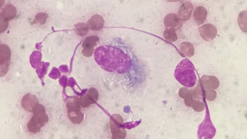
A 2yo MN Boxer presented for evaluation of multiple growths on the ears. They had appeared and progressed steadily over the last two weeks.
The dog was otherwise healthy and did not seem bothered by the growths. He was receiving an isoxazoline monthly and had no other major medical history.
Brittany Lancellotti, DVM, DACVD
Veterinary Skin and Ear, Los Angeles, CA
Exam:
On physical exam, the pet was bright, alert, responsive and euhydrated with a body condition score of 4/9. At the proximal spine of the helix and convex pinnae bilaterally, there were multifocal, alopecic, pink, dome-shaped, soft to firm nodules. The vertical and horizontal canals were nonerythematous, nonexudative, and nonedematous. Tympanic membranes were intact bilaterally. No excoriations or evidence of self-induced trauma from head shaking or ear scratching was noted. The remainder of the dermatologic exam was unremarkable.


Diagnostics:
Initial diagnostics included cytology, Wood’s lamp, skin scrape and fine needle aspirates.
Cytology from impression smear revealed primarily keratin debris with rare extracellular coccoid bacteria. No intact white blood cells were observed.
Woods lamp did not produce any fluorescence of the hair shafts on lesional skin.
Skin scrape was negative for Demodex and Sarcoptes mites.
Fine needle aspirate stained with Diff-Quik revealed granulomatous inflammation with intracellular negatively stained bacilli.


Search terms
Canine leproid granuloma, leproid granuloma boxer, canine leproid granuloma syndrome, disfiguring lesions, canine leprosy, most common mycobacterial disease, common mycobacterial disease, drug therapy, affected dogs