A 7 year 6-month-old, female spayed, Chinese Pug presents for a 4-month history of hair loss and pruritus. The owner reports she was first seen scratching at the axillary region, shoulders, and behind the ears.
Matthew Levinson, DVM, DACVD
Blue Pearl Pet Hospital, Northfield, IL
History:
Initially she was diagnosed with yeast infection in both ears secondary to suspected seasonal environmental allergies and was prescribed thiabendazole, dexamethasone, neomycin sulfate sodium topically for the yeast infection and hydroxyzine for itch relief. Despite these treatments the pruritus persisted and she was seen a month after initial presentation where she then received intramuscular dexamethasone injection and was sent home with oclacitinib 5.4mg daily dose. The pruritus continued five weeks after receiving the dexamethasone injection and being on oclacitinib for symptomatic relief, the primary veterinarian then prescribed oral amoxicillin trihydrate/clavulanate potassium, oral trimeprazine/prednisolone and Chlorhexidine 2%/ketoconazole 1% shampoo. There was mild improvement in terms of itch relief with the frequent bathing but after couple more visits to her primary veterinarian and no significant improvement the dog was referred to veterinary dermatologist. At the time, this dog presented for a dermatology consultation, she was on currently on hydroxyzine for itch relief and fipronil for flea and tick prevention. This was the only pet in the household.
Exam:
T: 102.8F
P: 120
mm: pink, moist
CRT: 1-2 seconds
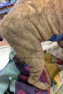
On physical exam, the patient had lichenification, hyperkeratotic crusting, and alopecia on the face, pinnae, neck, lateral thorax, flanks, limbs, and tail. There was hypotrichosis appreciated along the mid dorsum. The external ear canal had moderate ceruminous debris, discharge, and only partial visualization of the tympanic membranes were appreciated. The patient did have a positive pinnal-pedal reflex.


Figures 1-4: Marked hyperkeratotic crusting, alopecia, and lichenification seen to the face, pinnae, lateral thorax, flanks, and limbs
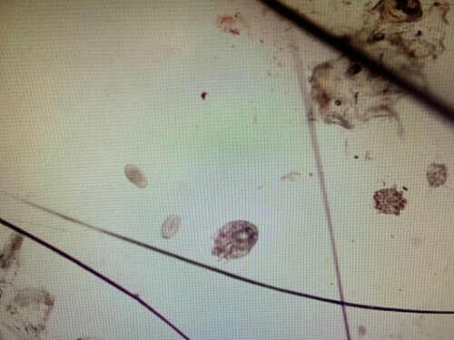
This image shows both eggs and adult Sarcoptic scabiei mites

Diagnostics:
Initial diagnostics included skin scraping and cytology.
Skin scraping revealed 2-10+ eggs, nymphs, and Sarcoptic scabiei adult mites per 4x field.
Cytology of the affected skin revealed corneocytes, nuclear streaming, pyogranulomatous
inflammation, too numerous to count cocci per oil immersion field
Cytology of both ears revealed proteinaceous debris with too numerous to count cocci and rods per oil immersion field
Assessment:
Sarcoptic mange
Secondary bacterial dermatitis
Bilateral otitis externa
Treatment plan:
References:
1.Miller WH, Griffin CE, Campbell KL. Autoimmune and immune-mediated dermatoses. In:
Muller and Kirk, eds. Small Animal Dermatology. 7th ed. St. Louis, MO: Elsevier; 2013: 315-316
2.Beugnet F, de Vos C, Liebenberg J, Halos L, Larsen D, Fourie J. 2016. Efficacy of afoxolaner in a clinical field study in dogs naturally infested with Sarcoptes scabiei. Parasite, 23, 26.
3.Pin D, Bensignor E, Carlotti DN, et al: Localised sarcoptic mange in dogs:
a retrospective study of 10 cases. J Small Anim Pract 47:611, 2006.
4.Bourdeau P, Armando L, Marchand A: Clinical and epidemiological characteristics
of 153 cases of sarcoptic acariosis in dogs. Vet Dermatol
15:48, 2004
5.Feather L, Gough K, Flynn RJ, et al: A retrospective investigation into risk
factors of sarcoptic mange in dogs. Parasitol Res 107:279, 2010.