


The skin, ear canal and gut of the dog harbor communities of bacteria and fungi whose balance contributes to the health of the host. In recent years, sequencing of microbial DNA has shown that these communities are far richer than culture had suggested.
This sequencing has revealed that these communities change in atopic dogs. Atopic skin loses diversity in favor of Staphylococcus pseudintermedius, which directly links the microbiome to flares and secondary infections. Malassezia pachydermatis is also significantly more abundant. Over the past ten years, interest has extended to the gut, because the intestinal microbiota contributes to the maturation of the immune system and could influence the skin at a distance. These data have given rise to new treatments, from probiotics to fecal microbiota transplantation. This review covers what canine studies establish, from the skin and ear to the gut, and what they still leave open.
1 Microbiota, microbiome, dysbiosis: definitions and methods
1.1 Definitions
The microbiota is the set of microorganisms living at a given site, and the microbiome the set of their genes. In practice, however, the two terms are often used interchangeably (Torres, 2017). Dysbiosis is an imbalance of this microbiota, quantitative or qualitative (Martin Cordero, 2019). On the skin, a resident population, relatively stable, is distinguished from a transient population that persists only for a few hours or days (Tizard, 2025). Both contain commensals and potential pathogens, so the presence of a microorganism is not enough to define a disease.
A probiotic is a live microorganism that confers a benefit on the host when consumed in sufficient quantity. A prebiotic is a non-digestible food component, often a fiber, that promotes the growth of beneficial bacteria. A postbiotic is a product of probiotic fermentation, including their metabolites and fragments of their cell wall. Finally, a synbiotic combines a probiotic and a prebiotic (Rostaher, 2026a). The international ISAPP definition also includes inactivated microorganisms and their components among postbiotics (Bonel-Ayuso et al., 2025). Bacteria are most often inactivated by heat, notably by tyndallization, which alternates temperatures of 70 to 100 °C with lower temperatures. The bacterium then keeps its cell wall, and its immunological effects remain close to those of the live bacterium (Santoro, 2022).
1.2 Methods and limitations of studies
Culture grows only a small proportion of the bacteria present. Sequencing, by contrast, identifies most of the genera present without culture: sequencing of the 16S ribosomal RNA gene for bacteria, and of the ITS region for fungi. This targeted sequencing, however, yields relative abundances: an increase in the proportion of staphylococci may reflect true overgrowth or a decline in other bacteria. Absolute abundances remove this ambiguity (Tang et al., 2020). Finally, metagenomics sequences all the DNA in the sample. It reaches species level, sometimes strain level, and describes gene functions (Whittle et al., 2024).
The skin carries a low quantity of microbes. A sample is therefore easily contaminated by reagents or by the environment. Sampling and analysis protocols vary from one team to another, which makes results difficult to compare. Sample sizes are often small, so a real difference may fail to reach statistical significance (Older and Rodrigues Hoffmann, 2024). Variability between individuals, breeds and sites adds to this. Finally, a cross-sectional study shows an association, never the direction of the relationship between dysbiosis and disease.
Laboratories now offer this sequencing as a diagnostic test. In 24 dogs with superficial bacterial folliculitis, two sequencing laboratories and one culture laboratory detected Staphylococcus equally often, with no difference in turnaround time. They agreed poorly, however, on methicillin resistance and on susceptibility to most antibiotics. Sequencing therefore does not yet replace culture for these lesions (Smart et al., 2025).
2 The skin microbiome: from the healthy dog to the atopic dog
2.1 In the healthy dog
Canine skin carries a rich microbiota that varies greatly from one individual to another. In a study of 12 healthy dogs, haired regions were richer and more diverse than mucosae and mucocutaneous junctions. The phylum Pseudomonadota (formerly Proteobacteria) dominated at all sites (Rodrigues Hoffmann et al., 2014). Within the same dog, haired skin keeps a similar composition from one site to another. The most frequent genera are Porphyromonas, Staphylococcus, Streptococcus, Cutibacterium and Corynebacterium, with large variations between studies (Rodrigues Hoffmann, 2022). In 6 healthy dogs sampled at eight sites, Bacillota (formerly Firmicutes) dominated, with a mean relative abundance of 34.4%. They were followed by Actinomycetota (formerly Actinobacteria), with 32.2%, Pseudomonadota, with 16.4%, and Bacteroidota (formerly Bacteroidetes), with 8.7% (Štempelová et al., 2025; Rodrigues Hoffmann, 2026). The dermis and subcutis, on the other hand, do not harbor a microbiota: in 7 healthy dogs, sequencing of biopsies did not differ from that of blank controls (García-Fonticoba et al., 2019). The fungi of healthy skin are mainly Alternaria and Cladosporium, coming from the environment, alongside variable proportions of Malassezia (Meason-Smith et al., 2015).
Several factors shape this composition. Breed and sampling site are the main ones in a metagenomic study of 72 dogs of four breeds (Whittle et al., 2024). In puppies, season, region, household hygiene and outdoor access modify the skin flora, which also differs from that of adults (Rodriguez-Campos et al., 2020). Dogs in the same household share a similar microbiota (Rodrigues Hoffmann, 2022). The skin microbiota also reflects the dog’s living environment and habits (Lehtimäki et al., 2018).
Despite this variability, a common core exists. In the 72 dogs of the metagenomic study, this core, known as the “core” microbiome, comprised 230 taxa and 1,219 gene families. It was also enriched in pathways for the synthesis of secondary metabolites. This richness suggests that the healthy microbiome protects against the establishment of pathogens, through a mechanism called colonization resistance (Whittle et al., 2024). Monthly follow-up of 15 healthy dogs for five months found coagulase-negative staphylococci, Sphingomonas, Malassezia, Vishniacozyma victoriae and Cladosporium in 70% of samples. None of these five organisms, however, was present in every dog at every visit (Harvey and Krumbeck, 2025). This core rebuilds itself after an insult. In 60 dogs, washing reduced the microbial load below the detection threshold, then the flora returned within 7 to 28 days through regrowth of taxa already present, with no input of new taxa (Whittle et al., 2026).
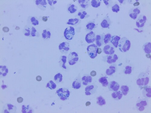
Images of phagocytosis of cocci in a dog
2.2 In the atopic dog
The skin of atopic dogs is less diverse than that of healthy dogs. As early as 2014, the 6 allergic dogs in a sequencing study had lower species richness than the 12 healthy dogs (Rodrigues Hoffmann et al., 2014). In 14 atopic dogs followed during a flare, bacterial diversity was reduced compared with 16 controls, in favor of Staphylococcus, especially S. pseudintermedius, and Corynebacterium (Bradley et al., 2016). A larger study, using 16S sequencing of the skin in 44 atopic dogs and 42 controls of three breeds, found enrichment in Staphylococcus at all sites and in all breeds, but also in Capnocytophaga, in bacteria of the Rhizobium group and in Aureimonas (Whittle et al., 2025).
Dysbiosis is most marked during flares. Outside flares and in the absence of infection, in 12 allergic German shepherd dogs, only the axilla was less rich than in 12 healthy German shepherds (Apostolopoulos et al., 2021). Similarly, in 3 atopic dogs without lesions, staphylococci were more abundant than in controls at each site, without the difference being significant. In the groin, for example, they reached 36.50% in atopic dogs versus 4.26% in controls (Chermprapai et al., 2019). In diseased dogs, dysbiosis is not limited to a change in proportions: bacterial and fungal biomass is much higher than on healthy skin (Tang et al., 2020).
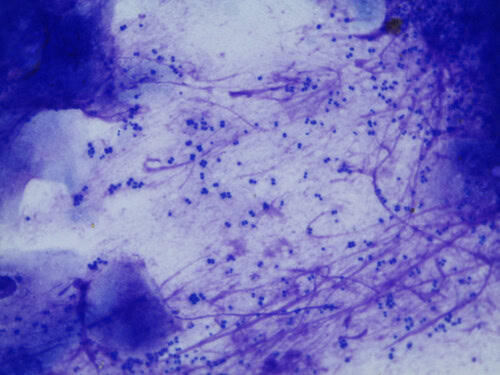
In atopic dogs, Staphylococcus pseudintermedius is the main species isolated
2.3 The mycobiome
The mycobiome, that is, the fungal population, also changes in allergic dogs. In 8 allergic dogs, it was less rich than in 10 healthy dogs. All sites also clustered according to clinical status (Meason-Smith et al., 2015). The change mainly concerns Malassezia species. Malassezia globosa was significantly more abundant on healthy skin than on the skin of allergic dogs, both by sequencing and by quantitative PCR. Malassezia restricta was also significantly more abundant there by sequencing. Malassezia pachydermatis, by contrast, was significantly more abundant, by sequencing, on the skin of naturally allergic dogs. In experimentally sensitized dogs, it was also more abundant, by quantitative PCR, in provoked lesions (Meason-Smith et al., 2020). Malassezia populations, however, did not change in step with the appearance of provoked lesions. It is therefore not known whether M. pachydermatis overgrowth precedes or follows the lesions. Commensal species and M. pachydermatis differ in their lipid dependence, which suggests a role for skin lipid composition in the community shift (Meason-Smith et al., 2020).
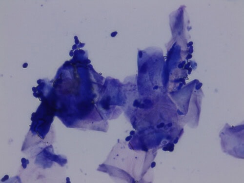
Malassezia pachydermatis is the yeast most frequently isolated in allergic dogs
2.4 Cause or consequence?
The experimental model shows that dysbiosis can follow allergy. In 8 dogs sensitized to house dust mites, a challenge on the right groin produced lesions in all dogs. At the challenged site, Corynebacteriaceae increased from the first day, then Staphylococcaceae from the seventh to the twenty-first day. Quantitative PCR confirmed an increase in S. pseudintermedius on days 7 and 21 compared with the opposite side (Pierezan et al., 2016).
Dysbiosis is also linked to the state of the skin barrier. In the 14 atopic dogs followed, bacterial diversity was correlated with transepidermal water loss and skin pH, but not with hydration measured by corneometry. Antimicrobial treatment restored diversity, reduced staphylococci and normalized transepidermal water loss and pH. Clinical severity decreased at the same time (Bradley et al., 2016). In humans, Th2 cytokines, in particular IL-4 and IL-13, inhibit skin defenses. Human dysbiosis is therefore both a consequence of inflammation and a factor that perpetuates it. In dogs, overgrowth of S. pseudintermedius likewise contributes to perpetuating inflammation (Mosca, 2026). Atopic skin also retains staphylococci more. In studies that still referred to Staphylococcus intermedius, bacteria adhered more to corneocytes of atopic dogs than to those of healthy dogs, and the more so as pruritus was intense (Simou et al., 2005). This increased adherence was observed on inflamed atopic skin as well as on normal-looking skin (McEwan et al., 2006). It depends mainly on the host: isolates from atopic and healthy dogs bound in the same way to matrix proteins (Schmidt et al., 2009). Likewise, corneocytes from persistent carrier dogs retained on average three times more bacteria than those of other dogs (Paul et al., 2013).
Conversely, dysbiosis can itself trigger inflammation. In mice lacking the protein ADAM17 in the epidermis, dysbiosis appeared spontaneously, with the successive emergence of Corynebacterium mastitidis, Staphylococcus aureus and Corynebacterium bovis. Antibiotics targeted at these bacteria almost completely abolished dysbiosis and skin inflammation. Staphylococcus aureus drove the development of eczema, whereas C. bovis induced a strong Th2 response (Kobayashi et al., 2015). This murine model has no equivalent in dogs.
The flora of the first weeks of life does not predict the disease. In a cohort of 143 West Highland white terriers, including 109 puppies aged three weeks or less, the early skin microbiota did not predispose to the later development of atopic dermatitis (Rodriguez-Campos et al., 2020). In dogs, the available data therefore place skin dysbiosis mainly downstream of inflammation, as an aggravating factor rather than as a primary cause.
2.5 From dysbiosis to superficial pyoderma
Superficial pyoderma lesions carry marked staphylococcal dysbiosis. In atopic dogs, 12 superficial bacterial folliculitis lesions and 9 epidermal collarettes were much less diverse than the skin of healthy dogs. Staphylococcus was more abundant there, especially in collarettes, and S. pseudintermedius was the main staphylococcal species in all sample types (Older et al., 2020).
3 The ear microbiome: from the healthy dog to otitis
3.1 The healthy ear
The cerumen of healthy dogs harbors a highly variable bacterial community, dominated by Pseudomonadota, Actinomycetota, Bacillota, Bacteroidota and Fusobacteria (Borriello et al., 2020). The healthy ear is slightly less diverse than the skin, and it carries low absolute quantities of microbial DNA. The most frequent organisms there are S. pseudintermedius, M. pachydermatis, Corynebacterium auriscanis and Enterococcus faecalis (Rodrigues Hoffmann, 2026). The fungal flora of the healthy ear is dominated by ascomycetes and includes environmental molds such as Alternaria, Cladosporium and Epicoccum (Prélaud, 2022).
3.2 The ear of the atopic dog without otitis
Dysbiosis affects the ear before any clinical otitis. In 11 atopic dogs without signs of otitis, the ear microbiota was imbalanced compared with 9 healthy dogs. Staphylococcus and Ralstonia were more abundant in atopic dogs, Escherichia in healthy dogs. This profile remained stable over twenty-eight days (Ngo et al., 2018). In allergic German shepherd dogs, the ear canal also differed from that of healthy German shepherds (Apostolopoulos et al., 2021). In allergic dogs, ear diversity decreases further during flares, in favor of Staphylococcus and Malassezia (Rodrigues Hoffmann, 2026).
3.3 Otitis externa
Otitis externa markedly reduces the diversity of the ear canal. Alpha diversity is the diversity within each sample. In 18 affected dogs, it was clearly reduced compared with 8 healthy dogs. The two groups differed by 153 taxonomic units (Korbelik et al., 2019). In 23 affected dogs, diversity also decreased, with an increase in Bacillota and Staphylococcus. The severity of otitis, however, did not change the profile (Kasai et al., 2021). Staphylococcus was the most abundant genus in 30 affected dogs as in 10 healthy dogs. Ears rich in cocci were the least diverse, and all forms of otitis were accompanied by reduced fungal diversity (Bradley et al., 2020). Of 129 samples from affected ears, 69.8% showed bacterial overgrowth, 16.3% fungal overgrowth and 7.0% both (Tang et al., 2020). A metagenomic study of 100 affected and 100 healthy dogs confirmed the predominant place of S. pseudintermedius in otitis externa: it accounted for 38.7% of relative abundance in affected ears. In healthy ears, Bacteroides fragilis dominated, with 15.8%, and Ezakiella coagulans, with 8.2% (Karakaya et al., 2026). Finally, in 6 fungal otitis cases, Malassezia accounted for 55.7 to 98.4% of fungal sequences, with a median of 96.8%. Fungal richness there was clearly lower than in 5 healthy ears (Korbelik et al., 2018).
3.4 Fungal dysbiosis after otic antibiotic therapy
Antibacterial treatment of otitis promotes Malassezia overgrowth, because it eliminates the bacteria that occupied the canal. In 25 dogs treated topically with a fluoroquinolone or with the combination of piperacillin and tazobactam, bacterial counts fell significantly, to a median of zero. But M. pachydermatis counts increased significantly in both groups. Candida was also isolated in 2 dogs after treatment (Juhola et al., 2023). In a second series of 15 dogs with Gram-negative bacterial otitis, adding clotrimazole reduced the risk of fungal dysbiosis: 6 of 15 dogs developed it, versus 14 of 14 in an earlier series treated with piperacillin and tazobactam alone (Gallow et al., 2025). Clotrimazole therefore reduces this dysbiosis without abolishing it (Gallow et al., 2025). After successful otic antibiotic therapy, yeast overgrowth should therefore be monitored (Juhola et al., 2023).
4 The gut-skin axis
4.1 Proposed mechanisms and canine data
The gut-skin axis refers to the influence of the intestinal microbiota on the skin. By breaking down fiber, intestinal bacteria produce short-chain fatty acids (SCFAs), which regulate immunity and strengthen the intestinal barrier. Intestinal dysbiosis could therefore act on the skin in several ways: through increased intestinal permeability, through the passage of metabolites or toxins into the circulation, through altered T-lymphocyte differentiation, or through reduced production of cutaneous antimicrobial peptides (Rodrigues Hoffmann, 2024). Three pathways are considered the main ones: SCFAs, tryptophan metabolites and microbial molecular patterns recognized by Toll-like receptors. These mechanisms have been demonstrated in basic research in other species, but the gut-skin axis remains poorly understood in dogs (Rostaher, 2026a).
Fecal concentrations of acetic, propionic and butyric acids were significantly lower in 25 atopic dogs than in 27 healthy dogs (Gonçalves et al., 2026). In 30 atopic dogs classified by severity, two markers of the intestinal wall decreased as the dermatitis worsened: diamine oxidase and zonulin. Zonulin thus fell from 14.67 to 10.89 ng/mL between mild and severe forms (Ural et al., 2026). In 8 dogs with both atopic dermatitis and adverse food reaction, compared with 9 controls, fecal bacterial diversity was reduced. The main SCFAs decreased, while lipid mediators derived from lipoxygenases increased (Hayashi et al., 2026). These three studies are pilot studies, without longitudinal follow-up.
4.2 The intestinal microbiota of the atopic dog
Studies comparing the intestinal microbiota of atopic and healthy dogs reach discordant results (Table 1). Some find lower diversity in atopic dogs, others similar diversity. One last study finds higher diversity, interpreted as instability of the microbiota (So Severo et al., 2026). The genera involved also vary from one study to another.
Table 1. Intestinal microbiota of atopic dogs compared with that of healthy dogs.
| Study | Population | Method | Main result |
|---|---|---|---|
| Rostaher et al., 2022 | 3 atopic beagles, 4 healthy | 16S | Lower diversity in atopic dogs; Lachnospiraceae, Anaerovoracaceae and Oscillospiraceae more abundant in healthy dogs; differences stable at one month |
| Uchiyama et al., 2022 | Shiba colony: 9 atopic, 16 healthy | 16S | Similar intestinal diversity; Anaerovoracaceae associated with dermatitis |
| Thomsen et al., 2023 | Atopic and healthy Shibas | 16S | Escherichia/Shigella and Clostridium sensu stricto abundant in atopic dogs; Fusobacteria and Megamonas reduced |
| Sugita et al., 2023 | 12 atopic, 20 healthy | 16S | Significantly different composition |
| Sinkko et al., 2023 | Privately owned Labradors and Finnish Lapphunds | Not specified | Different composition; severity linked to antibiotics received |
| Felten et al., 2025 | 12 allergic WHWTs, 9 healthy | Metagenomics | No difference; abundance of Escherichia coli inversely correlated with pruritus |
| Song et al., 2025 | 23 atopic, 11 healthy | 16S | Lower diversity; severity inversely correlated with diversity and with Fusobacterium and Megamonas |
| Whittle et al., 2025 | 44 atopic, 42 healthy, three breeds | Metagenomics | Enrichment in Escherichia, profile close to chronic enteropathy |
| So Severo et al., 2026 | 35 atopic, 15 healthy | 16S, rectal swab | Higher diversity in atopic dogs; Peptacetobacter hiranonis less frequent |
These discrepancies are partly due to the populations studied. Breed, diet, living environment and treatments received differ between studies, and sample sizes remain small. In Finnish dogs, diet was the factor most strongly linked to the intestinal microbiota, but it was only weakly linked to symptoms. Severity, for its part, was associated with antibiotics received. These antibiotics were themselves linked to the composition of the microbiota (Sinkko et al., 2023).
4.3 Diet and intestinal microbiota
Diet modifies the intestinal microbiota of allergic dogs. In 40 dogs fed for eight weeks a hydrolyzed diet based on fish and rice starch, SCFA-producing bacteria increased in all dogs. Dogs classified as atopic because they did not respond to the diet retained a profile different from that of dogs with food allergy, both before and after the elimination trial (Noli et al., 2023). In 32 atopic dogs, combining a nutraceutical with a hypoallergenic diet lowered the intestinal dysbiosis index as well as lesion and pruritus scores. This study, however, had no control group (Guidi et al., 2021).
5 The early-life window
5.1 Environment, lifestyle and the first months
The hygiene hypothesis and the biodiversity hypothesis link the decline in early microbial exposure to the rise in allergic diseases. They rest on the idea that diverse microbes “educate” the immune system during an early window of life (Rostaher, 2026a). In dogs, epidemiological data point in this direction. In a Finnish national survey of 5,722 dogs, allergic symptoms were more frequent in urban environments, in dogs as in their owners. A large family and regular contact with farm animals and other animals protected against these symptoms (Hakanen et al., 2018). This window opens at birth. In 18 French bulldog puppies born by cesarean section, the richness and diversity of the oral and rectal microbiota increased from birth to weaning. The skin of the dam’s mammary glands was their first source of microbes, before the maternal oral and rectal flora took over (Cicirelli et al., 2026). In 169 dogs of two breeds, allergy was most frequent in urban dogs living with a single person and with no contact with other animals. It was rarest in rural dogs with the opposite lifestyle (Lehtimäki et al., 2018). In 168 dog-owner pairs, urban living homogenized the skin microbiota of both species, which shared part of it. The bacteria associated with allergy nevertheless differed between dogs and humans (Lehtimäki et al., 2020).
Puppy diet adds to these factors. In a survey of 1,158 cases and 2,864 controls, feeding raw tripe or raw organ meats to puppies was associated with fewer allergic signs after one year: odds ratio of 0.36 for raw tripe and 0.23 for raw organ meats (Hemida et al., 2021). This study is based on questionnaires, so it shows an association and not a cause. Early use of antibiotics is also among the risk factors identified (Rostaher, 2026a).
5.2 Tolerance and regulatory T cells
Regulatory T cells (Tregs) ensure tolerance to environmental antigens and to commensals. In mice, a wave of Tregs reaches the skin just after birth and establishes tolerance to the skin flora (Rodrigues Hoffmann, 2024). In dogs, in a cohort of West Highland white terriers, the future healthy dogs had at three months a higher proportion of blood Tregs than the future atopic dogs: 4.5% versus 3.3% (Rostaher et al., 2021).
5.3 Early interventions in the experimental model
The high-IgE beagle model is based on a colony of beagles that produce large amounts of IgE. When sensitized to house dust mites through the skin, these dogs develop a dermatitis close to the natural disease clinically, histologically and immunologically (Marsella et al., 2006). This model made it possible to test an early intervention. An atopic bitch received Lactobacillus rhamnosus GG during her second pregnancy, then her puppies received it from three weeks to six months of age. The bitch’s two litters, the first untreated and the second treated, were then sensitized to Dermatophagoides farinae. At six months, the treated litter had lower specific IgE and weaker intradermal reactions than the untreated litter. Clinical scores did not differ between the two litters, and 6 of the 9 treated puppies nevertheless developed dermatitis after challenge (Marsella, 2009). Three years after the probiotic was stopped, clinical scores on challenge remained lower in the 9 exposed dogs than in the 7 unexposed dogs, whereas specific IgE no longer differed (Marsella et al., 2012). Early exposure did not change the expression of filaggrin, a barrier protein, in this model (Marsella et al., 2013).
Fecal microbiota transplantation has also been tried in puppies. Beagle puppies were sensitized to house dust mites after one of three treatments: none, metronidazole or fecal transplantation. Untreated puppies and puppies treated with the antibiotic had higher IgE and pruritus scores than transplanted puppies. These preliminary results, from unpublished work by Chapman et al., have only been presented at a congress (Rostaher, 2026a). Prevention of atopic dermatitis is therefore no longer a purely theoretical idea, but the evidence remains limited (Rostaher, 2026b).
6 Treatments and modulation of the microbiome
6.1 Impact of antimicrobials and washing
Studies on antibiotics remain short, and their long-term effects on the canine microbiome are not known (Rodrigues Hoffmann, 2022).
Antiseptic shampoos have a transient effect. In 3 atopic dogs and 6 healthy dogs, a shampoo containing 2% chlorhexidine and 2% miconazole, applied twice a week for three weeks, did not significantly change diversity. Bacterial diversity of atopic skin tended to increase during treatment, then returned to its initial level four weeks after treatment was stopped (Chermprapai et al., 2019). Repeated washing also modifies the flora of healthy dogs. In 16 Labradors washed daily for fourteen days with a diluted detergent, diversity increased, while the usually dominant phyla declined (Discepolo et al., 2023). In 20 atopic dogs with areas of dysbiosis, 3% chlorhexidine wipes, applied once a day for fourteen days, reduced clinical scores and cytological counts from the seventh day, without microbiome analysis or a control group (Bensignor et al., 2024).
In the ear, the choice of treatment modifies the microbiome. In 20 dogs with otitis externa, an antibiotic-free formulation and the combination of gentamicin, betamethasone and clotrimazole gave the same clinical cure. The conventional combination, however, led to overgrowth of Neisseria dumasiana, whereas the antibiotic-free formulation did not change any taxon (Charello Bannach et al., 2026). In 30 dogs with erythemato-ceruminous otitis associated with Malassezia, a ceruminolytic ear cleaner reduced pruritus by 77% and the clinical score by 66% in fourteen days. It increased fungal diversity without clearly changing the bacterial flora (Briand et al., 2025).
6.2 Impact of anti-inflammatory drugs
Six atopic dogs without lesions received ciclosporin for one month then prednisone for one month, separated by one month without treatment. Neither the diversity nor the composition of the bacterial microbiota, nor Malassezia populations, changed (Widmer et al., 2018). For oclacitinib, the two available studies diverge. In atopic Shibas, oclacitinib brought the composition of the skin and intestinal microbiota closer to that of healthy dogs (Thomsen et al., 2023). In 3 atopic beagles, four weeks of oclacitinib did not change the intestinal microbiota (Rostaher et al., 2022). In the anal sacs, the microbiota of 14 untreated atopic dogs differed from that of 15 healthy dogs. In 6 atopic dogs treated with oclacitinib, desloratadine or allergen-specific immunotherapy, by contrast, its structure was closer to that of healthy dogs (Bergeron et al., 2024).
Topical glucocorticoids do not unbalance the ear. In 10 atopic dogs without otitis, mometasone applied to a single ear for four weeks did not change bacterial or fungal diversity compared with the untreated ear, while clinical and cytological scores of the treated ear improved (Léonard et al., 2021). In 15 dogs with non-purulent otitis, prednisolone, combined with pomegranate in one group and with antimicrobials in the other, significantly reduced clinical signs on days 15 and 30. Fungal diversity increased after both treatments, through a decline in Malasseziaceae (Puigdemont et al., 2021; Rodrigues Hoffmann, 2026). In 5 beagles with mild erythematous otitis, a hydrocortisone aceponate spray reduced for fourteen days the genera associated with dysbiosis, notably Escherichia–Shigella, Moraxella and Macrococcus. Diversity then decreased transiently, before recovering after fourteen days without treatment (Ghibaudo et al., 2026). This last pilot study was funded by the manufacturer.
6.3 Oral pro-, post- and synbiotics
Trials of oral probiotics and postbiotics in atopic dogs are numerous, but small and heterogeneous (Table 2). Each trial tests a different strain, at a different dose and for a different duration, so their results cannot be added up.
Table 2. Clinical trials of oral probiotics, postbiotics and synbiotics in canine atopic dermatitis.
| Study | Product | Design | Number of dogs | Result |
|---|---|---|---|---|
| Kim et al., 2015 | Lactobacillus sakei Probio-65 | Versus placebo, 2 months | Not specified | Severity index reduced |
| Ohshima-Terada et al., 2015 | Lactobacillus paracasei K71 | Versus cetirizine, 12 weeks | 41 | Similar lesions and pruritus; medication score reduced |
| Osumi et al., 2019 | Inactivated Enterococcus faecalis FK-23 | Versus placebo, 84 days | 39 | CADESI-04 reduced; pruritus and medication unchanged |
| Yamazaki et al., 2019 | Enterococcus faecium SF68, with oclacitinib | Versus placebo, 12 weeks | 21 | No reduction in oclacitinib dose |
| Kawano et al., 2023 | L. paracasei M-1 and kestose (synbiotic) | Open-label, 90 days, dogs on prednisolone | 15 | Median CADLI from 28 to 12; median pruritus from 6 to 2; total prednisolone dose reduced |
| Tate et al., 2024 | Probiotics and nutraceuticals | Versus placebo, 10 weeks, pruritic dogs | 62 analyzed | Faster improvement; marked placebo effect |
| Cardoso et al., 2025 | L. rhamnosus GG, with oclacitinib | Versus placebo, 90 days | 40 | CADESI-04 reduced; pruritus improved in the treated group only |
| Song et al., 2025 | Three-strain probiotic | Open-label, no untreated atopic group, 16 weeks | 23 | Severity reduced; increase in intestinal diversity in good responders |
| Kiewsiri et al., 2026 | Inactivated Lactobacillus plantarum L-137, with oclacitinib | Crossover versus placebo, 12 weeks | 12 | Pruritus and CADESI-04 not significantly improved; serum CCL17 reduced; fecal microbiota unchanged |
Meta-analyses do not confirm a clear effect. A systematic review, limited to English-language articles from 2019 to 2023, included only 5 trials. In these trials, probiotics, oral or topical, did not significantly change either the CADESI-04, a score of the extent and severity of lesions, or the pruritus score (Pacheco et al., 2025). A meta-analysis of postbiotics in dogs, all indications combined, based on 13 in vivo studies, showed no significant difference from control groups on the criteria analyzed. The studies included had small sample sizes, heterogeneous strains and varied study designs (Bonel-Ayuso et al., 2025). An oral probiotic can nevertheless modify the skin at a distance: in 12 healthy golden retrievers, ninety days of supplementation reduced the frequency of S. pseudintermedius in the groin and increased bacterial diversity in the axilla (Grant et al., 2025).
6.4 Topical bacteria and targeted bacteriotherapy
In 10 mildly affected atopic dogs, a spray of heat-inactivated Lactobacillus rhamnosus and Lactobacillus reuteri was applied daily for twenty-eight days. It reduced the clinical score from the fourteenth day and pruritus on the forty-second day. The skin microbiota, however, had not changed at twenty-eight days (Santoro et al., 2021). A spray of two live strains, Lactiplantibacillus plantarum YUN-V2.0 and Lacticaseibacillus rhamnosus YUN-S1.0, reduced the clinical score in 7 mildly allergic dogs, without changing pruritus (Santoro, 2025; Mueller et al., 2026). In the ear, drops containing these two strains persisted for one week in the canal of healthy dogs and inhibited P. aeruginosa, S. pseudintermedius and M. pachydermatis in vitro (Claes et al., 2025). In 37 dogs with non-infected erythemato-ceruminous otitis, probiotic ear drops applied every other day for twenty-eight days lowered the mean lesion score from 5.11 to 1.66 and pruritus from 4.93 to 2.09. The trial was open-label, without a control group (Carrasco et al., 2026). These last three studies were conducted with the support of the manufacturer or by its employees.
Targeted bacteriotherapy looks for commensals that kill the pathogen. A strain of Staphylococcus felis isolated from cat skin, strain C4, secretes a bacteriocin, micrococcin P1, and antimicrobial peptides. In mice, it reduced skin colonization by methicillin-resistant S. pseudintermedius (MRSP), and an extract of its antimicrobials reduced the necrotic lesions it causes (O’Neill et al., 2021). On a canine skin explant, the live bacterium and its sterile protein product reduced the growth of a methicillin-susceptible strain and of a methicillin-resistant strain. Twenty-four hours after application of the sterile product, no organism was recovered (Faccin et al., 2025). A Lactobacillus casei isolated from the pinna of healthy dogs inhibited in vitro all 30 clinical isolates of S. pseudintermedius tested (Diniz et al., 2025). A bacteriophage specific to S. pseudintermedius, applied daily for thirty days, cured generalized superficial MRSP pyoderma in one dog (Mota Sá et al., 2025). These approaches remain at the laboratory or single-case stage.
6.5 Fecal microbiota transplantation
Fecal microbiota transplantation consists of providing the intestinal microbiota of a healthy donor. In an open-label trial, a single oral transplantation in 12 atopic dogs lowered the median CADESI-04 from 16.5 to 8 and the pruritus score from 3 to 1 in fifty-six days. It also changed the composition of the fecal microbiota (Sugita et al., 2023). A randomized, double-blind, placebo-controlled trial then enrolled 46 dogs. Of these, 40 completed the trial, 20 per group. The transplanted group received daily capsules of freeze-dried microbiota for ninety days and three monthly rectal administrations. CADESI-04 was lower with transplantation than with placebo at the second month (7 versus 16) and at the third (8 versus 15). Sustained responders were more frequent with transplantation: 35% versus 5% with placebo. They were defined by an improvement of at least 50% at both check-ups. At the third month, medication scores were 13 with transplantation versus 24 with placebo. Pruritus decreased in both groups, with no difference between them. Transplantation was well tolerated (Felten et al., 2026). This result, however, rests on a single controlled trial.
6.6 ICADA 2026 guidelines
The International Committee on Allergic Diseases of Animals (ICADA) included microbiome modulation in its 2026 guidelines. It grades the strength of its recommendations from A to D. Level A rests on consistent trials, level B on at least one randomized trial, level C on descriptive studies and level D on expert opinion. It retains two mechanisms, demonstrated in vitro: competition of beneficial bacteria with pathogens and the production of tolerogenic substances by host cells. The evidence is judged limited to recommend oral probiotics for the treatment or prevention of atopic dermatitis (strength B). Topical products containing live or tyndallized bacteria are considered safe and effective in mild forms (strength C), but there is no evidence that they modify the skin microbiome (strength B). The committee therefore proposes microbiome modulation as an option, during flares or as proactive maintenance in mildly affected dogs. The benefits of this modulation must nevertheless be weighed against inconsistent efficacy. It recalls that any recurrent infection is secondary to an underlying cause and that repeated courses of antibiotics or antifungals should be avoided (Mueller et al., 2026). The guidelines do not mention fecal transplantation, whose controlled trial was published the same year.
Conclusion
Cutaneous and otic dysbiosis in atopic dogs is now well established. It combines a loss of diversity, an overgrowth of S. pseudintermedius and M. pachydermatis, and an increase in microbial biomass. It follows allergic inflammation in the experimental model, worsens during flares and regresses with treatment. It is therefore a perpetuating factor rather than a demonstrated primary cause. The gut-skin axis, by contrast, remains a hypothesis in dogs, because studies of the intestinal microbiota contradict each other and the mechanisms have only been demonstrated in other species. Early-life factors (urban living, puppy diet, antibiotics) are associated with the risk of atopic dermatitis. Early exposure to probiotics had lasting effects in the beagle model, in the puppies of a single bitch. Therapeutically, oral probiotics have not demonstrated a clear effect. Topical bacteria improve mild forms without modifying the microbiome. Fecal transplantation reduced lesions and medication use in a single controlled trial. Current guidelines remain to treat infections while looking for their cause and to avoid repeated courses of antibiotics (Mueller et al., 2026).
Key take-home messages
- In the experimental model, skin dysbiosis can follow allergic inflammation; in atopic dogs, it is most marked during flares and regresses with antimicrobial treatment.
- A recurrent infection always has an underlying cause to be identified, and repeated courses of antibiotics or antifungals should be avoided.
- In atopic dogs, ciclosporin, prednisone and otic mometasone did not change the diversity of the microbiome.
- A topical antibiotic in the ear promotes the multiplication of Malassezia; combining it with clotrimazole limits this risk without eliminating it.
- A chlorhexidine and miconazole shampoo did not significantly change the diversity of the skin microbiome, which had returned to its baseline level four weeks after treatment was stopped.
- A few trials of oral probiotics are positive, but meta-analyses show no clear effect on lesions or pruritus.
- According to international guidelines, topical products containing bacteria are considered safe and effective in mild forms, but there is no evidence that they modify the skin microbiome.
- Fecal microbiota transplantation, using the feces of a healthy donor, reduced lesions and medication needs, with no difference in pruritus, in a single controlled trial.
- Urban living, few contacts with other people and other animals, and early antibiotics are associated with an increased risk of allergy, but the evidence that the disease can be prevented remains limited.
References
Apostolopoulos N, Glaeser SP, Bagwe R, Janssen S, Mayer U, Ewers C, et al. Description and comparison of the skin and ear canal microbiota of non-allergic and allergic German shepherd dogs using next generation sequencing. PLoS One. 2021;16(5):e0250695. doi:10.1371/journal.pone.0250695
Bensignor E, Navarro C, Gard C, Jahier B, Pressanti C, Videmont E. Efficacy of Chlorhexidine Impregnated Wipes for the Local Dysbiosis in Atopic Dogs: A Multicentric Prospective Study. Vet Sci. 2024;11(6). doi:10.3390/vetsci11060240
Bergeron C, Costa MC, Segura M, de Souza LB, Bleuzé M, Sauvé F. Bacterial microbiota and proinflammatory cytokines in the anal sacs of treated and untreated atopic dogs: Comparison with a healthy control group. PLoS One. 2024;19(5):e0298361. doi:10.1371/journal.pone.0298361
Bonel-Ayuso DP, Pineda-Pampliega J, Martinez-Alesón García P, Fernandez-Muela M, de la Fuente J, Garcia Fernandez PM, et al. Effects of Postbiotic Administration on Canine Health: A Systematic Review and Meta-Analysis. Microorganisms. 2025;13(7). doi:10.3390/microorganisms13071572
Borriello G, Paradiso R, Catozzi C, Brunetti R, Roccabianca P, Riccardi MG, et al. Cerumen microbial community shifts between healthy and otitis affected dogs. PLoS One. 2020;15(11):e0241447. doi:10.1371/journal.pone.0241447
Bradley CW, Lee FF, Rankin SC, Kalan LR, Horwinski J, Morris DO, et al. The otic microbiota and mycobiota in a referral population of dogs in eastern USA with otitis externa. Vet Dermatol. 2020;31(3):225-e49. doi:10.1111/vde.12826
Bradley CW, Morris DO, Rankin SC, Cain CL, Misic AM, Houser T, et al. Longitudinal Evaluation of the Skin Microbiome and Association with Microenvironment and Treatment in Canine Atopic Dermatitis. J Invest Dermatol. 2016;136(6):1182-1190. doi:10.1016/j.jid.2016.01.023
Briand A, Bensignor E, Dropsy H, Crosaz O, Humeau L, Cheval J, et al. Effect of a ceruminolytic ear cleaner on clinical, microbiological and ear canal microbiome evolution in canine erythemato-ceruminous otitis externa associated with proliferation of Malassezia yeasts. Vet Dermatol. 2025;36(5):668-678. doi:10.1111/vde.13352
Cardoso MC, Lopes TS, Lemos MC, Figueiredo CD, Streck AF. Efficacy of Lacticaseibacillus rhamnosus GG in the treatment of canine atopic dermatitis: A randomized controlled study. Braz J Microbiol. 2025;56(4):3081-3084. doi:10.1007/s42770-025-01767-x
Carrasco I, Homedes J, Arcas A, Beasley S, Salichs M. Clinical Efficacy of Probiotic-Based Ear Drops Containing Lactobacilli in Canine Erythroceruminous Otitis Externa: An Open Label, Multicentre Study. Vet Dermatol. 2026;37(5):818-826. doi:10.1111/vde.70097
Charello Bannach T, Mongruel A, Voi R, Farias M, Luciano F. Auricular microbiome responses to topical antibiotic-free versus conventional therapy in canine otitis externa. In: Proceedings of the 36th European Veterinary Dermatology Congress; 2026 Aug 27-29; Lille, France. p. 137.
Chermprapai S, Ederveen THA, Broere F, Broens EM, Schlotter YM, van Schalkwijk S, et al. The bacterial and fungal microbiome of the skin of healthy dogs and dogs with atopic dermatitis and the impact of topical antimicrobial therapy, an exploratory study. Vet Microbiol. 2019;229:90-99. doi:10.1016/j.vetmic.2018.12.022
Cicirelli V, Peruzzo A, Burgio M, Bramante G, Fabbri G, Toscan E, et al. Evaluation of neonatal oral and rectal microbiota in dogs delivered by c-section, from birth to weaning. Vet J. 2026;319:106816. doi:10.1016/j.tvjl.2026.106816
Claes I, Samp AK, Salichs M, Homedes J, Beasley S. Viability and pathogen inhibition activity of Lactiplantibacillus plantarum YUN-V2.0 and Lacticaseibacillus rhamnosus YUN-S1.0 present in a probiotic ear drops. In: Proceedings of the 35th European Veterinary Dermatology Congress; 2025 Sep 11-13; Bilbao, Spain. p. 137.
Diniz DCC, Ribeiro MG, Dias GS, Viana GB, Okamoto AS, Machado LHA. Antimicrobial activity of Lactobacillus casei on Staphylococcus pseudintermedius isolates. Vet Dermatol. 2025;36(4):485-493. doi:10.1111/vde.13325
Discepolo D, Kelley R, Watson A, Perry E. Impacts to canine dermal microbiota associated with repeated bathing. Front Vet Sci. 2023;10:1204159. doi:10.3389/fvets.2023.1204159
Faccin M, O’Neill AM, Lawhon SD, Worthing KA, Wiener DJ, Gallo RL, et al. Staphylococcus felis C4 exhibits in vitro antimicrobial activity against methicillin-resistant Staphylococcus pseudintermedius in a novel canine skin explant model. Vet Dermatol. 2025;36(1):24-33. doi:10.1111/vde.13308
Felten V, Turck JL, Unterer S, Favrot C, Suchodolski J, Fischer NM, et al. An insight into the gut microbiota of healthy and allergic West Highland Whiter Terrier dogs. PLoS One. 2025;20(8):e0328100. doi:10.1371/journal.pone.0328100
Felten V, West EA, Martini F, Favrot C, Unterer S, Suchodolski J, et al. Faecal Microbiota Transplantation Reduces Lesion Severity and Medication Use in Canine Atopic Dermatitis: A Randomised, Placebo-Controlled, Double-Blinded Clinical Trial. Vet Dermatol. 2026;37(5):735-744. doi:10.1111/vde.70092
Gallow G, Juhola J, Ferguson E, Loeffler A, Hendricks A, Frosini S, et al. Clotrimazole reduces but does not abolish fungal dysbiosis following potent antibacterial monotherapy for canine otitis externa. In: Proceedings of the 35th European Veterinary Dermatology Congress; 2025 Sep 11-13; Bilbao, Spain. p. 154.
García-Fonticoba R, Ferrer L, Francino O, Cuscó A. Dermis and subcutis of healthy dogs lack of a bacterial microbiota. In: Proceedings of the 31st European Veterinary Dermatology Congress; 2019 Sep 26-28; Liverpool, United Kingdom. p. 131.
Ghibaudo G, Olivero D, Gentilini F, Turba ME. Impact of a topical hydrocortisone aceponate spray on the canine ear canal microbiome: a pilot longitudinal study. In: Proceedings of the 36th European Veterinary Dermatology Congress; 2026 Aug 27-29; Lille, France. p. 152.
Gonçalves M, Fernandes B, Alves SP, Pereira H, Prego MT, Lourenço AM. Preliminary Measurement of Faecal Short-Chain Fatty Acids in Dogs With Canine Atopic Dermatitis. Vet Dermatol. 2026;37(1):45-50. doi:10.1111/vde.70015
Grant L, Mohammadi Dehcheshmeh M, Ebrahimie E, Khabiri A, Veltman T, Shipstone M, et al. The effect of daily oral probiotic and postbiotic supplementation on the canine skin microbiota: insights from culture-dependent and long-read 16S rRNA gene sequencing methods. In: Proceedings of the North American Veterinary Dermatology Forum; 2025 Apr. p. 164-165.
Guidi EEA, Gramenzi A, Persico P, Di Prinzio R, Di Simone D, Cornegliani L. Effects of Feeding a Hypoallergenic Diet with a Nutraceutical on Fecal Dysbiosis Index and Clinical Manifestations of Canine Atopic Dermatitis. Animals (Basel). 2021;11(10). doi:10.3390/ani11102985
Hakanen E, Lehtimäki J, Salmela E, Tiira K, Anturaniemi J, Hielm-Björkman A, et al. Urban environment predisposes dogs and their owners to allergic symptoms. Sci Rep. 2018;8(1):1585. doi:10.1038/s41598-018-19953-3
Harvey R, Krumbeck J. A 5-month temporal, quantitative study investigating the core microbiome on the axilla, umbilical region, and groin of 15 healthy dogs in the UK. Am J Vet Res. 2025;86(7). doi:10.2460/ajvr.25.01.0014
Hayashi A, Kakiyama S, Kawano K, Harima-Mizusawa N, Kobayashi T, Kaga C, et al. Fecal Lipid Metabolites and Microbiota Alterations in Dogs With Concurrent Atopic Dermatitis and Adverse Food Reaction. Allergy. 2026;81(7):2549-2552. doi:10.1111/all.70370
Hemida MBM, Salin S, Vuori KA, Moore R, Anturaniemi J, Rosendahl S, et al. Puppyhood diet as a factor in the development of owner-reported allergy/atopy skin signs in adult dogs in Finland. J Vet Intern Med. 2021;35(5):2374-2383. doi:10.1111/jvim.16211
Juhola J, Brennan E, Ferguson EA, Loeffler A, Hendricks A, Frosini SM, et al. Fungal dysbiosis following antibacterial monotherapy in canine otitis externa. In: Proceedings of the 34th European Veterinary Dermatology Congress; 2023 Aug 31-Sep 2; Gothenburg, Sweden. p. 156.
Karakaya E, Satıcıoğlu İB, Yarım D, Güran Ö, Güran C, Alpman U, et al. Culture and Metagenomic Insights into the Ear Microbiota in Dogs with Healthy Ears and Otitis Externa. Vet Sci. 2026;13(3). doi:10.3390/vetsci13030250
Kasai T, Fukui Y, Aoki K, Ishii Y, Tateda K. Changes in the ear canal microbiota of dogs with otitis externa. J Appl Microbiol. 2021;130(4):1084-1091. doi:10.1111/jam.14868
Kawano K, Iyori K, Kondo N, Yamakawa S, Fujii T, Funasaka K, et al. Clinical effects of combined Lactobacillus paracasei and kestose on canine atopic dermatitis. Pol J Vet Sci. 2023;26(1):131-136. doi:10.24425/pjvs.2023.145014
Kiewsiri C, Patumcharoenpol P, Kovitvadhi A, Chermprapai S. Efficacy of Heat-Killed Lactobacillus plantarum L-137 Supplementation in Treatment of Canine Atopic Dermatitis. Vet Dermatol. 2026. In press (published online). doi:10.1111/vde.70139
Kim H, Rather IA, Kim H, Kim S, Kim T, Jang J, et al. A Double-Blind, Placebo Controlled-Trial of a Probiotic Strain Lactobacillus sakei Probio-65 for the Prevention of Canine Atopic Dermatitis. J Microbiol Biotechnol. 2015;25(11):1966-9. doi:10.4014/jmb.1506.06065
Kobayashi T, Glatz M, Horiuchi K, Kawasaki H, Akiyama H, Kaplan DH, et al. Dysbiosis and Staphylococcus aureus colonization drives inflammation in atopic dermatitis. Immunity. 2015;42(4):756-766. doi:10.1016/j.immuni.2015.03.014
Korbelik J, Singh A, Rousseau J, Weese JS. Analysis of the otic mycobiota in dogs with otitis externa compared to healthy individuals. Vet Dermatol. 2018;29(5):417-e138. doi:10.1111/vde.12665
Korbelik J, Singh A, Rousseau J, Weese JS. Characterization of the otic bacterial microbiota in dogs with otitis externa compared to healthy individuals. Vet Dermatol. 2019;30(3):228-e70. doi:10.1111/vde.12734
Lehtimäki J, Sinkko H, Hielm-Björkman A, Laatikainen T, Ruokolainen L, Lohi H. Simultaneous allergic traits in dogs and their owners are associated with living environment, lifestyle and microbial exposures. Sci Rep. 2020;10(1):21954. doi:10.1038/s41598-020-79055-x
Lehtimäki J, Sinkko H, Hielm-Björkman A, Salmela E, Tiira K, Laatikainen T, et al. Skin microbiota and allergic symptoms associate with exposure to environmental microbes. Proc Natl Acad Sci U S A. 2018;115(19):4897-4902. doi:10.1073/pnas.1719785115
Léonard C, Taminiau B, Ngo J, Fantini O, Daube G, Fontaine J. Preventive use of a topical anti-inflammatory glucocorticoid in atopic dogs without clinical sign of otitis does not affect ear canal microbiota and mycobiota. Vet Dermatol. 2021;32(4):355-e98. doi:10.1111/vde.12977
Marsella R, Olivry T, Nicklin C, Lopez J. Pilot investigation of a model for canine atopic dermatitis: environmental house dust mite challenge of high-IgE-producing beagles, mite hypersensitive dogs with atopic dermatitis and normal dogs. Vet Dermatol. 2006;17(1):24-35. doi:10.1111/j.1365-3164.2005.00496.x
Marsella R, Santoro D, Ahrens K, Thomas AL. Investigation of the effect of probiotic exposure on filaggrin expression in an experimental model of canine atopic dermatitis. Vet Dermatol. 2013;24(2):260-e57. doi:10.1111/vde.12006
Marsella R, Santoro D, Ahrens K. Early exposure to probiotics in a canine model of atopic dermatitis has long-term clinical and immunological effects. Vet Immunol Immunopathol. 2012;146(2):185-9. doi:10.1016/j.vetimm.2012.02.013
Marsella R. Evaluation of Lactobacillus rhamnosus strain GG for the prevention of atopic dermatitis in dogs. Am J Vet Res. 2009;70(6):735-40. doi:10.2460/ajvr.70.6.735
Martin Cordero A. Bacterial skin infections in small animals. Zaragoza: Grupo Asís Biomedia; 2019.
McEwan NA, Mellor D, Kalna G. Adherence by Staphylococcus intermedius to canine corneocytes: a preliminary study comparing noninflamed and inflamed atopic canine skin. Vet Dermatol. 2006;17(2):151-154. doi:10.1111/j.1365-3164.2006.00503.x
Meason-Smith C, Diesel A, Patterson AP, Older CE, Mansell JM, Suchodolski JS, et al. What is living on your dog’s skin? Characterization of the canine cutaneous mycobiota and fungal dysbiosis in canine allergic dermatitis. FEMS Microbiol Ecol. 2015;91(12). doi:10.1093/femsec/fiv139
Meason-Smith C, Olivry T, Lawhon SD, Hoffmann AR. Malassezia species dysbiosis in natural and allergen-induced atopic dermatitis in dogs. Med Mycol. 2020;58(6):756-765. doi:10.1093/mmy/myz118
Mosca M. Biothérapies et inhibiteurs de JAK en dermatologie. In: VétoAlp 2026 : les défis de la dermatologie. AFVAC Rhône-Alpes; 2026 Mar 15-16; Chamonix-Mont-Blanc, France. p. 23-32.
Mota Sá C, Rodrigues C, Silva V, Azeredo J, Melo LDR. Successful bacteriophage therapy of pyoderma caused by methicillin-resistant Staphylococcus pseudintermedius in a dog. Vet Dermatol. 2025;36(2):227-230. doi:10.1111/vde.13328
Mueller RS, Banovic F, Bensignor E, Santoro D, Nuttall T. Treatment of Canine Atopic Dermatitis: Updated Guidelines From the International Committee on Allergic Diseases of Animals (ICADA). Vet Dermatol. 2026. In press (published online). doi:10.1111/vde.70128
Ngo J, Taminiau B, Fall PA, Daube G, Fontaine J. Ear canal microbiota – a comparison between healthy dogs and atopic dogs without clinical signs of otitis externa. Vet Dermatol. 2018;29(5):425-e140. doi:10.1111/vde.12674
Noli C, Varina A, Barbieri C, Pirola A, Olivero D. Analysis of Intestinal Microbiota and Metabolic Pathways before and after a 2-Month-Long Hydrolyzed Fish and Rice Starch Hypoallergenic Diet Trial in Pruritic Dogs. Vet Sci. 2023;10(7). doi:10.3390/vetsci10070478
O’Neill AM, Worthing KA, Kulkarni N, Li F, Nakatsuji T, McGrosso D, et al. Antimicrobials from a feline commensal bacterium inhibit skin infection by drug-resistant S. pseudintermedius. Elife. 2021;10:e66793. doi:10.7554/eLife.66793
Ohshima-Terada Y, Higuchi Y, Kumagai T, Hagihara A, Nagata M. Complementary effect of oral administration of Lactobacillus paracasei K71 on canine atopic dermatitis. Vet Dermatol. 2015;26(5):350-3, e74-5. doi:10.1111/vde.12224
Older CE, Rodrigues Hoffmann A, Hoover K, Banovic F. Characterization of Cutaneous Bacterial Microbiota from Superficial Pyoderma Forms in Atopic Dogs. Pathogens. 2020;9(8). doi:10.3390/pathogens9080638
Older CE, Rodrigues Hoffmann A. Considerations for performing companion animal skin microbiome studies. Vet Dermatol. 2024;35(4):367-374. doi:10.1111/vde.13250
Osumi T, Shimada T, Sakaguchi M, Tsujimoto H. A double-blind, placebo-controlled evaluation of orally administered heat-killed Enterococcus faecalis FK-23 preparation in atopic dogs. Vet Dermatol. 2019;30(2):127-e36. doi:10.1111/vde.12725
Pacheco RCF, Lins LF, DE Brito LP, DE Andrade Calaça PR, Porto ALF, Cavalcanti MTH. Probiotics as an adjunct in the treatment of canine atopic dermatitis: a systematic review and meta-analysis of in vivo studies in dogs. J Vet Med Sci. 2025;87(9):1068-1077. doi:10.1292/jvms.24-0471
Paul NC, Latronico F, Moodley A, Nielsen SS, Damborg P, Guardabassi L. In vitro adherence of Staphylococcus pseudintermedius to canine corneocytes is influenced by colonization status of corneocyte donors. Vet Res. 2013;44(1):52. doi:10.1186/1297-9716-44-52
Pierezan F, Olivry T, Paps JS, Lawhon SD, Wu J, Steiner JM, et al. The skin microbiome in allergen-induced canine atopic dermatitis. Vet Dermatol. 2016;27(5):332-e82. doi:10.1111/vde.12366
Prélaud P. Otites et dermatite atopique canine. In: Journées annuelles du GEDAC (AFVAC); 2022. p. 21.
Puigdemont A, D’Andreano S, Ramió-Lluch L, Cuscó A, Francino O, Brazis P. Effect of an anti-inflammatory pomegranate otic treatment on the clinical evolution and microbiota profile of dogs with otitis externa. Vet Dermatol. 2021;32(2):158-e37. doi:10.1111/vde.12930
Rodrigues Hoffmann A, Patterson AP, Diesel A, Lawhon SD, Ly HJ, Elkins Stephenson C, et al. The skin microbiome in healthy and allergic dogs. PLoS One. 2014;9(1):e83197. doi:10.1371/journal.pone.0083197
Rodrigues Hoffmann A. Gut microbiome and atopic dermatitis in people and animals. In: Proceedings of the 10th World Congress of Veterinary Dermatology; 2024 Jul; Boston, USA. p. 137-140.
Rodrigues Hoffmann A. Otic microbiome: from healthy ears to otitis externa and media. In: Proceedings of the North American Veterinary Dermatology Forum; 2026 Apr 30-May 2. p. 121-124.
Rodrigues Hoffmann A. The skin microbiome of dogs and cats. In: Proceedings of the North American Veterinary Dermatology Forum; 2022 Apr. p. 100-105.
Rodriguez-Campos S, Rostaher A, Zwickl L, Fischer N, Brodard I, Vidal S, et al. Impact of the early-life skin microbiota on the development of canine atopic dermatitis in a high-risk breed birth cohort. Sci Rep. 2020;10(1):1044. doi:10.1038/s41598-020-57798-x
Rostaher A, Morsy Y, Favrot C, Unterer S, Schnyder M, Scharl M, et al. Comparison of the Gut Microbiome between Atopic and Healthy Dogs-Preliminary Data. Animals (Basel). 2022;12(18). doi:10.3390/ani12182377
Rostaher A, Rodriguez-Campos S, Deplazes P, Zwickl L, Akdis AC, Urwyler A, et al. Atopic dermatitis in West Highland white terriers – Part III: early life peripheral blood regulatory T cells are reduced in atopic dermatitis. Vet Dermatol. 2021;32(3):239-e63. doi:10.1111/vde.12939
Rostaher A. Prevention of canine atopic dermatitis – turning dreams into reality. In: Proceedings of the 36th European Veterinary Dermatology Congress; 2026b Aug 27-29; Lille, France. p. 35-36.
Rostaher A. Targeting the microbiome for canine atopic dermatitis treatment. In: VétoAlp 2026 : les défis de la dermatologie. AFVAC Rhône-Alpes; 2026a Mar 15-16; Chamonix-Mont-Blanc, France. p. 14-17.
Santoro D, Fagman L, Zhang Y, Fahong Y. Clinical efficacy of spray-based heat-treated lactobacilli in canine atopic dermatitis: a preliminary, open-label, uncontrolled study. Vet Dermatol. 2021;32(2):114-e23. doi:10.1111/vde.12915
Santoro D. Beneficial bacteria and atopic dermatitis: how do they interact with the immune system? In: Proceedings of the North American Veterinary Dermatology Forum; 2022 Apr. p. 92-100.
Santoro D. Clinical efficacy of spray containing two strains of Lactobacilli in canine allergic dermatitis: a preliminary, open-label, uncontrolled study. In: Proceedings of the 35th European Veterinary Dermatology Congress; 2025 Sep 11-13; Bilbao, Spain. p. 202.
Schmidt V, Nuttall T, Fazakerley J, McEwan N. Staphylococcus intermedius binding to immobilized fibrinogen, fibronectin and cytokeratin in vitro. Vet Dermatol. 2009;20(5-6):502-508. doi:10.1111/j.1365-3164.2009.00804.x
Simou C, Thoday KL, Forsythe PJ, Hill PB. Adherence of Staphylococcus intermedius to corneocytes of healthy and atopic dogs: effect of pyoderma, pruritus score, treatment and gender. Vet Dermatol. 2005;16(6):385-391. doi:10.1111/j.1365-3164.2005.00484.x
Sinkko H, Lehtimäki J, Lohi H, Ruokolainen L, Hielm-Björkman A. Distinct healthy and atopic canine gut microbiota is influenced by diet and antibiotics. R Soc Open Sci. 2023;10(4):221104. doi:10.1098/rsos.221104
Smart K, Pieper JB, Viall AK, Noxon JO, Berger DJ. Comparison of commercial next-generation sequencing assays to conventional culture methods for bacterial identification and antimicrobial susceptibility of samples obtained from clinical cases of canine superficial bacterial folliculitis. Vet Dermatol. 2025;36(1):14-23. doi:10.1111/vde.13299
So Severo J, Mendes R, Gmyterco V, Farias M, Mongruel A, Fischer N, et al. Assessment of the gut microbiome and its relationship with the severity of atopic dermatitis in dogs. In: Proceedings of the 36th European Veterinary Dermatology Congress; 2026 Aug 27-29; Lille, France. p. 193.
Song H, Mun SH, Han DW, Kang JH, An JU, Hwang CY, et al. Probiotics ameliorate atopic dermatitis by modulating the dysbiosis of the gut microbiota in dogs. BMC Microbiol. 2025;25(1):228. doi:10.1186/s12866-025-03924-6
Štempelová L, Micenková L, Andrla P, Strompfová V. The skin microbiome on healthy and inflammatory altered canine skin determined by next generation sequencing. Front Microbiol. 2025;16:1528747. doi:10.3389/fmicb.2025.1528747
Sugita K, Shima A, Takahashi K, Ishihara G, Kawano K, Ohmori K. Pilot evaluation of a single oral fecal microbiota transplantation for canine atopic dermatitis. Sci Rep. 2023;13(1):8824. doi:10.1038/s41598-023-35565-y
Tang S, Prem A, Tjokrosurjo J, Sary M, Van Bel MA, Rodrigues-Hoffmann A, et al. The canine skin and ear microbiome: A comprehensive survey of pathogens implicated in canine skin and ear infections using a novel next-generation-sequencing-based assay. Vet Microbiol. 2020;247:108764. doi:10.1016/j.vetmic.2020.108764
Tate DE, Tanprasertsuk J, Jones RB, Maughan H, Chakrabarti A, Khafipour E, et al. A Randomized Controlled Trial to Evaluate the Impact of a Novel Probiotic and Nutraceutical Supplement on Pruritic Dermatitis and the Gut Microbiota in Privately Owned Dogs. Animals (Basel). 2024;14(3). doi:10.3390/ani14030453
Thomsen M, Künstner A, Wohlers I, Olbrich M, Lenfers T, Osumi T, et al. A comprehensive analysis of gut and skin microbiota in canine atopic dermatitis in Shiba Inu dogs. Microbiome. 2023;11(1):232. doi:10.1186/s40168-023-01671-2
Tizard IR. Veterinary immunology. 11th ed. St. Louis: Elsevier; 2025.
Torres S. Skin microbiome: the little we know about this fascinating microscope world. In: Proceedings of the North American Veterinary Dermatology Forum; 2017 Apr. p. 49.
Uchiyama J, Osumi T, Mizukami K, Fukuyama T, Shima A, Unno A, et al. Characterization of the oral and faecal microbiota associated with atopic dermatitis in dogs selected from a purebred Shiba Inu colony. Lett Appl Microbiol. 2022;75(6):1607-1616. doi:10.1111/lam.13828
Ural K, Erdoğan S, Erdoğan H, Pasa S, Özalp T. Leaky gut and intestinal mucosal injury among dogs with clinically subclassified atopic dermatitis. Braz J Vet Med. 2026;48:e011725. doi:10.29374/2527-2179.bjvm011725
Whittle M, Carvell-Miller L, Abrams C, Watson P, Amos G. Disruption, recovery, and stability of the canine skin microbiome after chlorhexidine and mild detergent intervention. In: Proceedings of the 36th European Veterinary Dermatology Congress; 2026 Aug 27-29; Lille, France. p. 211.
Whittle M, Gilroy R, Castillo-Fernandez J, McKay L, Brenda F, Chodroff Foran R, et al. Uncovering microbiome biomarkers of canine atopic dermatitis. In: Proceedings of the 35th European Veterinary Dermatology Congress; 2025 Sep 11-13; Bilbao, Spain. p. 218.
Whittle MJ, Castillo-Fernandez J, Amos GCA, Watson P. Metagenomic characterisation of canine skin reveals a core healthy skin microbiome. Sci Rep. 2024;14(1):20104. doi:10.1038/s41598-024-63999-5
Widmer G, Ferrer L, Favrot C, Paps J, Huynh K, Olivry T. Glucocorticosteroids and ciclosporin do not significantly impact canine cutaneous microbiota. BMC Vet Res. 2018;14(1):51. doi:10.1186/s12917-018-1370-y
Yamazaki C, Rosenkrantz W, Griffin C. Pilot evaluation of Enterococcus faecium SF68 as adjunctive therapy for oclacitinib-responsive adult atopic dermatitis in dogs. J Small Anim Pract. 2019;60(8):499-506. doi:10.1111/jsap.13042