


A fifteen-year-old male neutered domestic long-haired cat presented with a roughly two-year history of generalized pruritus, traumatic lesions, and alopecia. The generalized pruritus had been treated with barrier clothing, oral systemic steroids, and Atopica. The pet parent reported that the patient has not been able to be medicated successfully, is generally comfortable although historically the pruritus had waxed and waned.
Meghan Solc, DVM, DACVD
Dermatology for Animals, Catonsville, Maryland
August 2026
History
The patient presented to the dermatology practice with patches of alopecia and focal ulcerations. He was not on any consistent medications although Atopica was still being given on occasion. The lesions had been present for approximately 2 years with no historical pruritus or lesions. Previous labwork had been performed and was unremarkable other than 1 year previously where a general leukopenia characterized by a mild neutrophilia was noted. This has subsequently been normal upon repeat labwork.
General Exam
- Temperature: 101.3 Degrees Fahrenheit
- Respiration: 40 breaths per min
- Pulse: 200 beats per min
- Mucous membranes: pale pink and moist
- Capillary refill time: less than 2 sec
- Weight: 4.04 kg
- Thoracic Evaluation revealed clear lung sounds and a grade II-III/VI heart murmur
Dermatologic Exam
- Dull, short orange hair coat with patches of alopecia
- Four patches of alopecia with fine silver scale and few focal areas of ulcerations
- No oral lesions
- No pinnal or paw pad lesions
- Waxing and waning overgrooming and pruritus
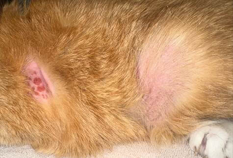
Figure 1: Photo of skin lesions
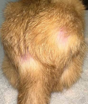
Figure 2: Photo of skin lesions
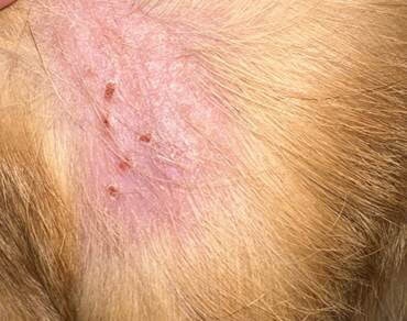
Figure 3: Photo of Lesions
Assessment
The lesions were concerning based on the patient’s history. Cytology and subsequent biopsy were recommended.
Diagnostics
- Cytology of the lesions revealed numerous rafts of round cells
- Histopathology: cutaneous lymphoid proliferation compatible with either cutaneous lymphocytosis or cutaneous small cell lymphoma
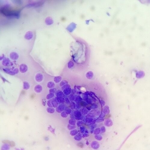
Figure 4: Cytology of Lesions
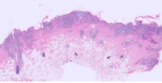
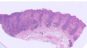
Figure 5: Histopathology of Lesions – Low Power
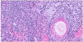
Figure 6: Histopathology of Lesions
Treatment Plan
- Dexamethasone orally 0.2mg/kg at a tapering schedule
- Referral to oncology
Follow-up
At the oncology referral and evaluation, the recommendation for further testing was made. PCR for antigen receptor rearrangements (PARR) testing and immunohistopathology was recommended. PARR testing was performed. Immunohistochemical staining was not pursued due to cost.
PARR testing was sent to Colorado State University and was consistent with a clonal population of T-lymphocytes. Based on these PARR results a small cell indolent T-cell lymphoma was considered more likely.
Treatment options are being considered. Specifically, chlorambucil and lomustine are options for treatment in addition to systemic steroid. The choice of treatment will be dependent on ease of administration and tolerance.
Discussion
Cutaneous small cell indolent T-cell lymphoma is a rare dermatologic diagnosis in cats. Most cats will develop lesions later in life and pet parents may not pursue full testing. Testing typically includes surface cytology and skin biopsy including PARR and immunohistochemistry tests.
There doesn’t appear to be a breed or sex predisposition although it is most common in older cats. Unlike in dogs, the adnexal structures are not as commonly affected. Range of survival time is variable and is difficult to predict due to the lower number of cases that are reported. The prognosis following diagnosis can range a few months up to 4 years.
Key Take Aways
- Lymphocytosis or cutaneous T-cell lymphoma should be a differential for all older cats with focal lesions
- Lesions can be progressive with or without pruritus
- Perform surface cytology to look for atypical cells
- Biopsy should always be considered in a cat with atypical lesions or abnormal cytology
References
Fontaine, J., Heimann, M, Day, M.J. Cutaneous epitheliotropic T-cell Lymphoma in the cat: a review of the literature and five new cases. Veterinary Dermatology. 2011 Oct; 22 (5):454-61.
Quintavalla, F., DiLecce, R. et al. Multifocal cutaneous non-epitheliotropic B-cell lymphoma in a cat. Journal of Feline Medicine and Surgery Open Reports. 2020