


“Teddy” is a 10 year old male neutered cocker spaniel presented by the owner for multiple cutaneous nodules of 3 months’ duration. He has a chronic history of pancreatitis well managed with a prescription low-fat diet. Both eyes were enucleated 3 years prior to presentation for the skin due to complications related to progressive glaucoma and cataracts.
Andrew Simpson
Animal Dermatology Clinic
Wheaton
August 2026
Subjective
History
In addition, “Teddy” has a history of chronic otitis externa managed with dexamethasone-based otic solution instilled in the ear canals once weekly along with weekly ear flushes at home.
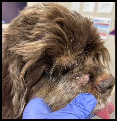
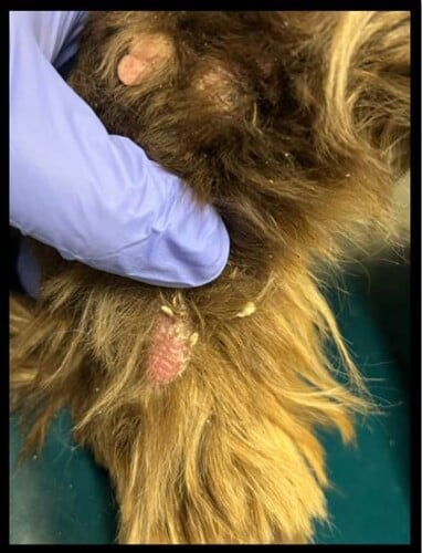
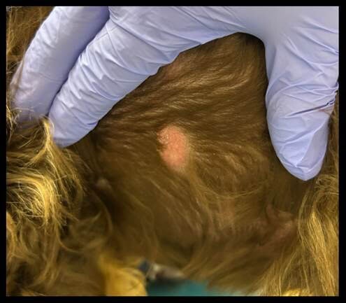
The owner reports that “Teddy” is not bothered by the nodules showing no signs of pruritus, exudate, or hemorrhage. The first nodules appeared on the dorsal head, now spreading to the extremities and along the entire dorsum and flanks.
His appetite and water consumption are reported as normal, with no changes in his activity level.
Current medications:
- Simparica (sarolaner) given by mouth once monthly year-round for flea and tick control
- Heartgard Plus (ivermectin + pyrantel pamoate) given by mouth once monthly year-round for heartworm and intestinal parasite prevention.
- Dexamethasone 4 mg/ml + saline otic solution (2:1 ratio): 0.4 ml in both ear canals twice weekly
Objective
Body Weight: 16.2 kg
Temperature: 100.1 F
Pulse: 115 bpm
Respiratory: 38 bpm
Bright, alert, responsive
General examination
no lesions on examination of oral cavity; heart and lungs auscult clearly in all fields with no arrhythmias noted; Prominent submandibular lymph nodes bilaterally. Mild enlargement of prescapular and popliteal lymph nodes; abdomen is soft and non-painful on palpation; no neurologic abnormalities noted.
Dermatologic examination
1) Multiple rounded to oval-shaped erythematous plaquiform to nodular lesions located in multiple areas, measuring approximately 1 cm on the right side of the muzzle, the left tarsal region, as well as multiple nodular-like lesions present on the dorsal aspect of multiple digits, especially the front paws.
Video otoscopic examination findings
– Right ear: No active inflammation present. No stenosis or masses noted. The tympanic membrane is fully visible and appears within normal limits.
– Left ear: Mild stenosis with moderate ceruminous gland hyperplasia along the vertical and horizontal aspect of the canal. There is a moderate amount of yellow cerumen present at the entrance of the canal. The tympanic membrane is fully visible and appears within normal limits.
Differential diagnoses for multiple cutaneous nodules:
- Inflammatory: sterile granuloma/pyogranuloma syndrome, cutaneous reactive histiocytosis, cutaneous plasmacytosis
- Infectious: fungal (blastomycosis, histoplasmosis, coccidioidomycosis), bacterial (deep staphylococcal pyoderma, Mycobacteria, Nocardia)
- Neoplastic: cutaneous lymphoma, mast cells tumors, plasmacytomas, histiocytic sarcomas, carcinomas.
- Other: nevi/hamartomas
Diagnostics
- Fine needle aspiration of nodules: poor exfoliation, non-diagnostic
- Fine needle aspiration of popliteal and submandibular lymph nodes: reactive lymphadenopathy, no neoplastic cells or infectious organisms noted
- Ear swab cytology (both ears): no infectious organisms or inflammatory cells present.
- Complete blood cell count: no significant findings
- Serum biochemistry: moderate hyperglobulinemia 5.9 (2.4 – 4.0 g/dL)
- Urinalysis: no significant findings
- Skin biopsy of 3 cutaneous nodules (muzzle, left tarsus, dorsum):
In all samples, the dermis, deep dermis, and subcutis are expanded to varying degrees by a population of monomorphic plasma cells arranged in sheets. Nuclei are eccentrically placed and are irregularly round with stippled chromatin and small nucleoli. The cells form nodular masses. Mitoses are rare. Low numbers of binucleate and multinucleate cells are noted. Scattered well-differentiated inflammatory mast cells are noted. The epidermis is mildly hyperplastic and there is orthokeratotic to parakeratotic hyperkeratosis.
Assessment
Diagnosis: Cutaneous plasmacytosis
The skin biopsy results were supportive of cutaneous plasmacytosis. Based on the presence of plasma cell disease in addition to hyperglobulinemia found on bloodwork, it was recommended to pursue diagnostic imaging as well as serum electrophoresis and screening for Bence-Jones proteins out of concern for multiple myeloma.
Follow-up Diagnostics
- Thoracic radiographs: normal cardiopulmonary structures, no lymphadenopathy noted
- Abdominal ultrasound: normal examination, no significant findings
- Serum protein electrophoresis: polyclonal gammopathy
- Bence-Jones protein (urine): none detected
Based on these diagnostic results, multiple myeloma was deemed less likely due to the lack of systemic involvement. In “Teddy’s” case, it appears that the multiple plasmacytomas (cutaneous plasmacytosis) were confined to the skin and likely the cause of the hyperglobulinemia.
Treatment and Outcome
The owner declined treatment with conventional chemotherapy after consulting with a boarded veterinary oncologist. Surgical resection of the growths was deemed impractical due to the disseminated distribution of nodules.
Prednisone was chosen as treatment to help reduce the number and size of the nodules (1 mg/kg/day by mouth once daily, tapered to every other day after 14 days). The prednisone slightly reduced the size of the cutaneous nodules, but did not resolve them. Instead of pursuing other therapies, the owners elected to monitor. There has been no progression of disease at the 4 month follow-up examination.
Discussion
Cutaneous plasmacytosis in dogs is a rare neoplasm involving multiple plasmacytomas confined to the skin, with no signs of multiple myeloma systemically.1 Canine plasma cell tumors (plasmacytomas) more commonly present as solitary tumors on the skin in which adequate surgical margins provide a cure. They appear as pink, raised, alopecia, well-circumscribed nodules 1-2 cm in diameter on the face or limbs more commonly.2 In one study, multiple cutaneous plasma cell tumors were reported relatively rarely, in 2.2% of 406 dogs.3
This is in contrast with true multiple myeloma in dogs, which involves neoplastic plasma cells in the bone marrow. Visceral organ involvement may also occur, including liver, spleen, kidneys, and abdominal lymph nodes.
There are very few reports in the veterinary literature describing multiple plasmacytomas with one retrospective case series published.1 Based on this study, the more common breeds reported include golden retrievers and Labrador retrievers. Breed disposition for solitary plasma cell tumors (plasmacytomas) is reported in American cocker spaniels, English cocker spaniels, and West Highland white terriers.1,2
It is not clear how many cases with multiple myeloma will develop cutaneous disease, however, in the retrospective case series involving dogs with cutaneous plasmacytosis, none of the dogs were reported to later develop multiple myeloma. Polyclonal gammopathy is uncommon in dogs with cutaneous plasmacytosis,1 although the dog in this report did have this finding.
Diagnosis requires skin biopsy, with possible immunohistochemistry staining to identify plasma cells as the origin. It would be advised to screen for systemic involvement in cases with multiple cutaneous plasma cell tumors. These diagnostics include: thoracic radiographs, abdominal ultrasound with aspiration and cytologic evaluation of the liver and spleen, survey radiographs of long bones (to screen for lytic bone lesions), serum protein electrophoresis, and urinalysis to evaluate Bence-Jones proteins.
Treatment can involve surgical resection when feasible (<5 cutaneous masses), though these cases may still develop additional plasmacytomas in the future. Several chemotherapy drugs were described in the study, however, melphalan provided the best overall response rate and the longest progression-free interval.1 Overall, the median survival time from the first treatment was 542 days.
References
Boostrom BO, Moore AS, DeRegis CJ, Robat C, Freeman K, Thamm DH. Canine Cutaneous Plasmacytosis: 21 Cases (2005-2015). J Vet Intern Med. 2017 Jul;31(4):1074-1080.
Lucke VM. Primary cutaneous plasmacytomas in the dog and cat. J Small Anim Pract 1987;28:49–55.
Goldschmidt MH, Shofer FS. Skin Tumors of the Dog and Cat. Oxford: Pergamon Press; 1992:265–270.