Color dilution alopecia (CDA), also known as color mutant alopecia, is a canine genodermatosis characterized by progressive hair loss specifically affecting areas of the coat with diluted pigmentation. We are pleased to present a comprehensive synthesis of knowledge about this dermatosis.
1. Introduction
Color dilution alopecia (CDA), also known as color mutant alopecia, is a canine genodermatosis characterized by progressive hair loss specifically affecting areas of the coat with diluted pigmentation. Although considered relatively uncommon in the overall canine population, it represents the most commonly diagnosed hereditary dermatosis in dogs. Its recognition and thorough understanding are fundamental for the veterinary dermatologist. They allow not only establishing a definitive diagnosis and distinguishing it from other alopecic conditions, but also providing informed and precise counsel to concerned owners and breeders. The disease manifests as a degradation of coat quality, evolving toward often extensive alopecia, and can be accompanied by secondary skin lesions, particularly bacterial infections.
The complexity of CDA lies not only in its clinical manifestations but also in its terminology and relationships with other follicular dysplasias. Historically, various terms have been used, and its distinction from black hair follicular dysplasia (BHFD) has been a subject of discussion. BHFD, which selectively affects areas of black coat in dogs with white spotting, presents notable histopathological similarities with CDA. This relationship suggests a common pathogenic basis, where a primary abnormality of pigmentation and follicular structure would be central. This terminological and classificatory nuance reflects the evolution of knowledge and underscores the importance of a rigorous diagnostic approach for these genetically determined dermatological conditions.
2. Etiopathogenesis of CDA
The etiopathogenesis of color dilution alopecia is multifactorial, involving specific genetic bases, complex pathophysiological mechanisms at the level of the hair follicle and pigmentation, as well as factors that modulate its clinical expression.
2.1. Genetic Basis: The MLPH Gene and its Variants
The genetic foundation of CDA rests predominantly on mutations within the melanophilin (MLPH) gene. This gene plays a crucial role in coding for an essential protein for the transport and distribution of melanosomes, the organelles containing melanin, within melanocytes and toward surrounding keratinocytes. The dilution of coat color, a phenotypic prerequisite for the development of CDA, is the direct consequence of these mutations.
Several recessive allelic variants (‘d’) of the MLPH gene have been identified as responsible for this dilution phenotype. The most extensively studied and frequently implicated variant is a single nucleotide polymorphism (SNP) located at position c.-22 relative to the translation initiation site in exon 1 of the MLPH gene, consisting of a substitution of a guanine (G) with an adenine (A) (c.-22G>A). Studies have confirmed a close association between this SNP and the dilute coat phenotype in many canine breeds. Besides this main variant (often designated d1), other mutations within the MLPH gene have been described, notably the d2 variant, a c.705G>C substitution identified in the Chow Chow, and the d3 variant, a c.667_668insC insertion reported in the Chihuahua. For a dog to phenotypically express a dilute coat color, and thus be predisposed to CDA, it must be homozygous for one of these recessive alleles (d/d genotype).
CDA is recognized as a disease with autosomal recessive transmission. This mode of transmission implies that both parents of an affected animal must be, at minimum, heterozygous carriers of the mutated allele (D/d) or be themselves affected (d/d).
2.2. Pathophysiological Mechanisms
Mutations of the MLPH gene induce a cascade of cellular and tissue events leading to the clinical manifestations of CDA.
Disruption of melanosome transport and aggregation: The main functional consequence of MLPH mutations is a defect in the melanosome transport mechanism. This results in an accumulation and anarchic aggregation of these pigmentary organelles, forming large inclusions called macromelanosomes, within the melanocytes of the epidermis and hair follicles, as well as in the keratinocytes of developing hair shafts. The d/d variant of MLPH is directly responsible for this defect in the homogeneous dispersion of melanosomes, leading to their aggregation. The presence of these macromelanosomes constitutes a distinctive histopathological and trichoscopic characteristic of CDA.
Follicular dysplasia and alteration of hair structure: The intracytoplasmic accumulation of these macromelanosomes, coupled with possible cytotoxicity exerted by melanin precursors or by the abnormal melanosomes themselves on the hair matrix cells, is considered a major factor in the development of follicular dysplasia. Hair follicles then become structurally abnormal, presenting distortions, progressive atrophy, and disturbances in the hair cycle. Simultaneously, the very structure of the hair shafts is compromised. The disorderly integration of macromelanosomes into the cortex and medulla of the hair alters its biomechanical integrity, making it abnormally fragile, brittle, and prone to premature fractures. This hair fragility is a direct contributor to the observed alopecia.
Keratinization abnormalities: Keratinization disorders frequently accompany CDA. Hyperkeratosis, particularly at the follicular level, is a common observation, manifesting as the formation of keratin plugs obstructing the hair infundibula. Excessive desquamation of the skin surface (scales) is also often reported. These keratinization abnormalities contribute to the dry and rough appearance of the skin and may favor infectious complications.
2.3. Genetic Complexity and Factors Influencing Expression
A fundamental and perplexing aspect of CDA is that the presence of a MLPH d/d genotype, while necessary, is not systematically sufficient to induce the alopecic phenotype. Indeed, not all dogs homozygous recessive for the dilution variants develop CDA, or they develop it with varying severity and age of onset. This phenomenon, known as incomplete penetrance, strongly suggests the involvement of other genetic factors (modifier genes) or environmental factors in the clinical expression of the disease. For example, it is well established that genetic tests for MLPH variants identify the coat dilution status but cannot predict whether a dilute-colored dog will actually develop CDA. The incidence of CDA in blue Dobermans is very high, but does not reach 100%, while in other breeds such as the Italian Greyhound, it is significantly lower despite the presence of dilute coats. Even more strikingly, certain breeds such as the Weimaraner or the Great Dane, which can present dilute coats (and thus a d/d genotype), rarely, if ever, manifest the clinical signs of CDA.
The existence of modifier genes is therefore a predominant hypothesis to explain this inter- and intra-breed variability. Genes such as RAB27A and MYO5A, which code for proteins functionally interacting with melanophilin within the melanosome transport complex, have logically been evoked as potential candidates. However, their direct and specific role in modulating the severity of CDA in d/d dogs has not yet been formally demonstrated by targeted studies. Research has indicated that the risk of developing CDA or BHFD seems to be breed-specific, which reinforces the idea that the overall genetic background of each breed plays a determining modulatory role. The precise identification of these modifier factors, whether genetic or environmental, remains an active and crucial field of research. The older hypothesis of a ‘dl’ allele at the D locus, recessive to ‘d’ and directly responsible for the alopecia, is less emphasized in recent publications, which tend more toward the interaction between MLPH and distinct modifier genes.
This evolution of understanding, from a strict monogenic model to a polygenic or multifactorial model, has considerable implications. In terms of genetic counseling, this means that an MLPH test alone, while informative about dilution status, is insufficient to predict with certainty the risk of developing alopecia. For research, this underscores the need for more global approaches, such as genome-wide association studies (GWAS) or whole-genome sequencing in cohorts of affected and unaffected d/d dogs, to identify these elusive modifier genes. Eventually, the characterization of these factors could not only refine prognosis but also open the way to new preventive or therapeutic strategies, if these modulators proved to be accessible targets. The identification of these elements could explain why certain lineages or breeds are more vulnerable than others, even when sharing the same MLPH d/d genotype.
3. Epidemiological Aspects
The study of the distribution and determinants of CDA within canine populations provides valuable information for its recognition and management.
3.1. Prevalence and Incidence
Color dilution alopecia is generally considered a relatively uncommon condition if we consider the entire canine population. Nevertheless, within the group of genodermatoses, it stands out for its higher diagnostic frequency, positioning it as the most commonly identified hereditary skin disease in dogs. Precise epidemiological data concerning its prevalence or incidence on the scale of large and diverse canine populations remain limited. However, the scientific literature, rich in individual case studies and clinical case series reports, attests to its presence in a wide range of canine breeds and in various geographical regions around the world.
3.2. Predisposed Breeds
A marked breed predisposition is a salient epidemiological characteristic of CDA. It mainly affects breeds in which coats with diluted pigmentation – such as blue (dilution of black), fawn or isabella (dilution of brown/chocolate), or lilac – are not only recognized by breed standards but sometimes actively sought after by breeders and owners. The following table synthesizes information relating to the breeds most frequently reported as being predisposed to CDA.
Table 1: Breeds Predisposed to Color Dilution Alopecia (CDA) and Frequency
|
Breed |
Concerned Dilute Colors |
Reported Frequency of CDA (if available) |
|---|---|---|
|
Doberman Pinscher |
Blue, Fawn (Isabella) |
Very high: 57.9% to 93% in blues; 75% to 89.5% in fawns. |
|
Dachshund |
Blue, Isabella |
Frequently reported. |
|
Yorkshire Terrier |
Blue (on blue and tan coat) |
Frequently reported, affecting the blue areas of the coat. |
|
Miniature Pinscher |
Blue, Fawn |
Reported. |
|
Whippet |
Blue, Fawn |
Frequently reported. |
|
Italian Greyhound |
Blue, Fawn |
Reported; incidence seems lower than in the Doberman despite the frequency of dilute coats. |
|
Chow Chow |
Blue |
Reported. |
|
Weimaraner |
Mouse-grey (Isabella) |
Genetically d/d, but clinical CDA is rarely observed, strongly suggesting the action of protective modifier genes. |
|
Great Dane |
Blue |
Similar situation to the Weimaraner; blue coats exist, but clinical CDA is infrequent. |
|
Labrador Retriever |
Charcoal (Blue), Silver (Lilac) |
CDA is increasingly recognized in these non-standard dilution colors. |
|
French Bulldog |
Blue |
CDA is reported in blue-colored individuals. |
|
American Staffordshire Terrier |
Blue |
Recognized predisposition. |
|
Other breeds |
Various dilute colors |
Standard Poodle (blue), Irish Setter (fawn), Saluki, Bernese Mountain Dog, Chihuahua, Shetland Sheepdog, Boston Terrier, Newfoundland, Schipperke, Beauceron, German Shepherd, Blue Gascony Hound. |
|
Mixed breed dogs |
Dilute coats of various origins |
CDA is not exclusive to purebred dogs and has been diagnosed in mixed breed dogs with dilute coats. |
This list is not exhaustive, but it illustrates the wide distribution of predisposition to CDA among canine breeds. The variability of clinical expression, even within predisposed breeds and for the same MLPH d/d genotype (such as the case of the Weimaraner), is a strong argument in favor of the influence of modifier genetic factors or poorly characterized environmental factors.
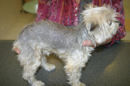
Severe color dilution alopecia in a Yorkshire
3.3. Influence of Age and Sex
The age of onset of the first clinical signs of CDA is variable but generally falls within a window ranging from 4 months to 3 years. Cases have been reported with symptom onset as early as 3 months of age, and more rarely, in older animals, with a case having been described in a 10-year-old dog. This wide range of age of onset, including within the same breed, could reflect the heterogeneity of modifier factors influencing the expression of the disease, or the interaction with environmental triggers yet to be identified. If the pathogenesis were solely determined by the MLPH d/d genotype, one might expect a more narrowed window of onset, unless gene expression is subject to intrinsically variable developmental regulation. Thus, this temporal variability, like incomplete penetrance, suggests an etiological complexity where additional genetic, epigenetic, or environmental factors (such as follicular stress related to grooming, nutrition, or general hormonal state) could interact with the primary genetic predisposition to modulate the timing of clinical manifestation.
Regarding the influence of sex, the majority of studies and clinical observations do not report a significant sexual predisposition for CDA. Males and females seem to be affected with comparable frequency, which is consistent with the autosomal recessive mode of transmission of the disease, independent of sex chromosomes.
4. Clinical Manifestations
The clinical manifestations of color dilution alopecia are progressive and characteristic, affecting the quality and density of the coat as well as the integrity of the underlying skin.
4.1. Age of Onset and Evolution
Typically, puppies destined to develop CDA are born with a normal-appearing coat in areas of dilute color. The first signs of the disease manifest insidiously and progressively, most often between the ages of 4 months and 3 years, although variations exist as mentioned previously. The alopecia is characterized by its slow progression, often extending over several months to several years. It can evolve to near-total hair loss on the trunk regions with diluted pigmentation.
4.2. Description of Hair and Skin Lesions
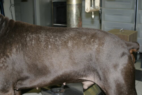
Coat quality: The first clinical indicator is frequently an alteration in the quality of the coat in areas of dilute color. The hair loses its luster, becoming dull, dry, and brittle. Affected hairs may present a “moth-eaten” appearance or diffuse thinning, leading to hypotrichosis. The remaining hair shafts are often rough to the touch and break easily under the effect of minor trauma such as rubbing or brushing.
Alopecia: The initial hypotrichosis inevitably evolves toward more marked alopecia, whose extent and symmetry can vary. The first areas affected are classically located on the trunk, particularly along the dorsal line. The alopecia can then extend to the flanks and the abdominal region. Characteristically, the head, limbs, and tail are often spared or are affected only late in the evolution of the disease. A pathognomonic sign, in the presence of a dilute coat, is the strict sparing of non-dilute colored areas. For example, in a blue and tan Doberman Pinscher, the tan markings will not be affected by the alopecia.
Primary and secondary skin lesions: In addition to hair loss, various skin lesions can be observed:
- Follicular papules and pustules are frequently encountered. These can evolve toward the formation of comedones (blackheads), resulting from the obstruction of hair follicles.
- Desquamation, presenting as fine (pityriasiform) or larger (furfuraceous) scales, is a common manifestation, reflecting keratinization disorders.
- Secondary bacterial pyoderma is a very frequent and clinically significant complication. It generally manifests as folliculitis or furunculosis, most often caused by the proliferation of Staphylococcus pseudintermedius. This bacterial superinfection is often the main cause of pruritus in dogs affected with CDA.
- Hypomelanotic macules, that is, areas of skin showing depigmentation, can appear, particularly on the ventral surface of the abdomen.
- Pruritus (itching) is typically absent or minimal in the absence of infectious complications. When present, it is almost systematically attributable to secondary bacterial pyoderma.
Mottled appearance of color dilution alopecia in a Dobermann
The sequence of appearance of these lesions constitutes an important element for diagnostic orientation. The initial modifications of coat quality, followed by progressive hypotrichosis then alopecia, generally precede the development of cutaneous complications such as pyoderma. This chronology, associated with the characteristic distribution of lesions (trunk mainly affected, sparing of non-dilute areas), is highly suggestive of CDA in a dog presenting a coat with diluted pigmentation. This helps to differentiate CDA from other dermatological conditions whose onset would be more abrupt or whose inflammatory signs would be primary.
5. Diagnostic Approach
The diagnosis of color dilution alopecia relies on a methodical approach integrating data from the history, clinical signs, and results of specific complementary examinations, notably trichogram and histopathological examination of skin biopsies.
5.1. History and Clinical Examination
A detailed history is the first crucial step. It should aim to gather precise information concerning the dog’s breed, coat color (and confirmation of the presence of pigmentary dilution), the exact age of onset of the first dermatological signs, the nature and chronology of the evolution of the lesions, as well as any family history (presence of other affected dogs in the same lineage or litter). Previous treatments and the observed response should also be documented.
The general and dermatological clinical examination must be exhaustive. It will confirm the presence of a dilute coat (blue, fawn, isabella, etc.) and allow precise characterization of the nature, distribution, and severity of the alopecic lesions, as well as the possible presence of primary or secondary skin lesions (papules, pustules, comedones, scales, erythema, crusts, hypomelanotic macules), in accordance with the descriptions provided in the previous section. The evaluation of the extent of alopecia and the search for signs of secondary pyoderma are particularly important.
5.2. Contribution of the Trichogram
The microscopic examination of hairs (trichogram) is a non-invasive diagnostic tool, simple to perform, quick, and of great informative value in the suspicion of CDA. Hairs are collected by gentle traction in the alopecic areas or on the periphery of the lesions, mounted between slide and coverslip in mineral oil or lactophenol, and observed under an optical microscope.
Typical observations in CDA include:
- The presence of numerous and voluminous melanin aggregates, or macromelanosomes, of irregular shape and size, distributed anarchically within the cortex and medulla of the hair shafts. These macromelanosomes are the microscopic signature of the pigmentary distribution abnormality.
- These pigmentary accumulations can lead to significant distortion of the hair shaft architecture, making it irregular, and cause points of weakness leading to transverse or oblique fractures of the hair.
- Other structural abnormalities of hairs can be visualized, such as deformed, twisted hair shafts, presenting abnormal angulations, or a damaged or absent cuticle in places, especially at the level of macromelanosome concentrations.
5.3. Skin Biopsy and Key Histopathological Features
Skin biopsy, followed by histopathological examination by an experienced veterinary pathologist, is often the examination of choice to confirm the diagnosis of CDA, particularly in atypical cases or to exclude other dermatoses. It is recommended to perform several biopsies (6 or 8 mm punch) on representative areas of the alopecia, including if possible primary lesions such as follicular papules, as well as an area of apparently healthy skin for comparative purposes.
The histopathological features considered typical and diagnostic of CDA are summarized in Table 2.
Table 2: Main Histopathological Features of Color Dilution Alopecia (CDA)
|
Histopathological Feature |
Preferential Location |
Detailed Description |
Diagnostic Importance |
|---|---|---|---|
|
Melanin Aggregates (Macromelanosomes) |
Epidermis (basal layer), Follicular epithelium (sheaths, matrix), Hair shafts, Dermis (melanophages) |
Large granulations or blocks of melanic pigment, of irregular shape and size, often massive. |
Very high; quasi-pathognomonic sign in compatible clinical context. |
|
Follicular Hyperkeratosis |
Infundibulum of hair follicles |
Thickening of the corneal layer within the follicle, leading to the formation of lamellar keratin plugs that dilate the follicular opening. |
High; contributes to comedones and folliculitis. |
|
Follicular Dysplasia |
Hair follicles (whole structure) |
Follicles of irregular shape, twisted, distorted, sometimes atrophic, cystic, or presenting a “witches’ foot” appearance. Numerous follicles in telogen phase. |
High; reflects the developmental and functional abnormality of the follicle. |
|
Pigmentary Incontinence / Dermal Melanophages |
Dermis (mainly peribulbar and perifollicular) |
Presence of free melanic pigment in the dermis and/or macrophages (melanophages) having phagocytosed this pigment, following its release by damaged epidermal or follicular cells. |
Moderate to high; indicates pigmentary remodeling. |
|
Epidermal Hyperkeratosis |
Interfollicular epidermis |
Thickening of the corneal layer of the epidermis, often of orthokeratotic type. |
Moderate; contributes to the scaly appearance of the skin. |
|
Follicular Atrophy |
Hair follicles |
Reduction in hair follicle size, particularly in advanced stages. |
Moderate; consequence of chronic dysplasia. |
|
Inflammatory Infiltrate |
Follicular, Perifollicular, Dermal |
In the absence of superinfection, the infiltrate is generally minimal to slight, lymphoplasmacytic. In the case of secondary pyoderma, a neutrophilic (suppurative) or pyogranulomatous (in the case of furunculosis) infiltrate will be observed. |
Variable; depends on the presence of complications. |
The combination of these lesions, in particular the presence of macromelanosomes in a context of follicular dysplasia and hyperkeratosis in a dog with a dilute coat, is strongly suggestive of the diagnosis of CDA.
5.4. Differential Diagnosis
It is imperative to distinguish CDA from other canine dermatological conditions that can manifest as alopecia, whether localized or generalized. Table 3 presents the main differential diagnoses and the criteria for distinguishing them from CDA.
Table 3: Differential Diagnosis of Color Dilution Alopecia (CDA) and Distinctive Criteria
|
Condition |
Key Clinical Signs (besides alopecia) |
Typical Trichogram Results |
Typical Histopathology Results |
Specific Complementary Tests |
|---|---|---|---|---|
|
Color Dilution Alopecia (CDA) |
Dilute coat, progressive alopecia on trunk, scales, follicular papules/pustules, frequent secondary pyoderma, pruritus if infection. Sparing of non-dilute areas. |
Macromelanosomes, deformed/fractured hair shafts. |
Follicular hyperkeratosis, macromelanosomes (epidermis, follicles, hairs), follicular dysplasia, dermal melanophages. |
History, clinical, trichogram, biopsy. MLPH genetic test (predisposition). |
|
Hypothyroidism |
Symmetrical, non-pruritic alopecia (unless pyoderma), dull, dry coat, thickened skin (myxedema), lethargy, weight gain. |
Hairs mostly in telogen, no macromelanosomes. |
Follicular atrophy, epidermal and follicular hyperkeratosis, dermal mucinosis. Absence of macromelanosomes. |
Total T4, TSH, free T4 (by equilibrium dialysis) measurement. |
|
Hypercorticicism (Cushing’s Syndrome) |
Bilateral symmetrical truncal alopecia, thin, atrophic skin, comedones, cutaneous calcinosis, abdominal distension, polyuria-polydipsia. |
Hairs mostly in telogen, no macromelanosomes. |
Epidermal, dermal, and follicular atrophy, follicular hyperkeratosis, comedones, calcium deposits (calcinosis). Absence of macromelanosomes. |
Low-dose dexamethasone suppression tests, ACTH stimulation test, urinary cortisol/creatinine ratio. |
|
Black Hair Follicular Dysplasia (BHFD) |
Alopecia affecting only areas of black coat in dogs with white spotting. Early onset. |
Similar to CDA but only on black hairs: macromelanosomes, structural abnormalities. |
Almost identical to CDA: macromelanosomes in black areas, follicular dysplasia. |
Clinical (distribution of lesions), trichogram, biopsy. |
|
Alopecia X (Nordic breed follicular dysplasia) |
Progressive symmetrical alopecia (trunk, neck, thighs), cutaneous hyperpigmentation. Predisposed breeds (Spitz, Husky, Malamute). |
“Flame-shaped” hairs (catagen/telogen), no macromelanosomes. |
Follicular atrophy, infundibular hyperkeratosis, trichilemmal keratinization. Absence of macromelanosomes. |
Exclusion of endocrinopathies, biopsy. Sometimes responds to castration or melatonin. |
|
Generalized Demodicosis |
Multifocal to generalized alopecia, erythema, scales, comedones, papules, pustules, variable pruritus. |
Visualization of Demodex canis (adults, larvae, eggs). |
Suppurative or granulomatous folliculitis/perifolliculitis with presence of demodex in the follicles. |
Deep skin scrapings, trichogram, (biopsy if scrapings negative and strong suspicion). |
|
Dermatophytosis |
Circular alopecic lesions, scaly, sometimes crusty, variable pruritus. Can be generalized. |
Fungal spores and/or hyphae on or in hair shafts (direct examination with KOH or lactophenol). |
Folliculitis/perifolliculitis, perivascular dermatitis. Visualization of fungal elements with special stains (PAS, Grocott). |
Wood’s lamp examination (fluorescence for certain strains of Microsporum canis), fungal culture on Sabouraud or DTM medium. |
5.5. Genetic Tests
Molecular genetic tests are commercially available to identify the different variants (‘d’ alleles, including d1, d2, d3) of the MLPH gene that are responsible for the dilution of coat color. These tests allow determination of whether a dog possesses the recessive homozygous genotype (d/d) necessary to express a dilute coat and, consequently, to be genetically predisposed to CDA.
However, it is crucial to emphasize that, although these tests confirm an animal’s dilution status, they cannot predict with absolute certainty whether a dog carrying the d/d genotype will actually develop the clinical signs of alopecia. This limitation is directly related to the phenomenon of incomplete penetrance of CDA and the probable influence of modifier genes or environmental factors, as discussed in section 2.3.
Nevertheless, these genetic tests are of considerable importance for breeding programs. They allow the identification of heterozygous dogs (D/d), which possess a non-dilute coat phenotype (unless other dilution genes are present) but are carriers of the ‘d’ allele and can therefore transmit it to their offspring. By avoiding matings between two carrier dogs (D/d x D/d) or between a carrier and a d/d individual, breeders can significantly reduce, or even eliminate, the risk of producing d/d puppies likely to develop CDA.
The diagnostic approach to CDA is therefore an integrative process that cannot rely on a single examination. It combines rigorous analysis of the signalment and clinical signs, the results of the trichogram, and the conclusions of the histopathological examination. The genetic test for the MLPH gene fits into this approach as a valuable tool to confirm genetic predisposition to dilution and to guide breeding advice. However, the diagnosis of the clinical disease, that is, the expression of alopecia, remains dependent on the confrontation of all clinical and paraclinical data. Future identification of more specific biomarkers or genetic tests targeting the modifier genes of CDA would constitute a major diagnostic and prognostic advance.
6. Therapeutic Management and Prognosis
The management of color dilution alopecia is primarily symptomatic, given the absence of a curative treatment for this genetic condition.
6.1. General Management Principles
To date, there is no treatment capable of correcting the underlying genetic defect of CDA or restoring a normal follicular structure and function in areas of dilute coat. The structural abnormalities of hair follicles and hair shafts induced by the pigmentation defect are considered permanent.
Consequently, the therapeutic management of CDA aims at several objectives:
- Managing associated cutaneous symptoms, such as dryness, desquamation, and pruritus (if present).
- Improving, as far as possible, the quality of the skin and remaining coat.
- Actively preventing and effectively treating secondary skin infections, particularly bacterial pyodermas.
- Maintaining a good quality of life for the affected dog.
6.2. Symptomatic Treatments
Topical care: Regular use of adapted topical products is the cornerstone of CDA management.
- Keratomodulating and emollient shampoos: These shampoos are used to control the keratoseborrheic state (desquamation, excess sebum or dryness) and to maintain adequate hydration of the skin and coat. Active ingredients such as benzoyl peroxide can be useful for their degreasing, antibacterial, and comedolytic action, particularly in the presence of folliculitis or comedones. Other agents such as sulfur, salicylic acid, or various moisturizing and emollient agents (urea, glycerol, colloidal oatmeal, vegetable oils) can also be beneficial depending on the specific clinical picture.
- Antiseptic shampoos: The use of shampoos containing antiseptic agents, such as chlorhexidine (at concentrations of 2% to 4%), is essential for the long-term management and prevention of secondary bacterial pyodermas, which are a frequent complication. The frequency of antiseptic baths must be adapted to the severity of the infections and can vary from once to several times a week in the acute phase, then be spaced out in the maintenance phase.
Management of bacterial superinfections: In the case of proven bacterial pyoderma (folliculitis, furunculosis), particularly if it is deep, extensive, or recurrent, systemic antibiotic therapy is often indispensable. First-line antibiotics, such as cephalexin or amoxicillin-clavulanic acid combination, are generally prescribed for a duration of 3 to 6 weeks, or at least one week after the clinical resolution of the lesions. In cases of chronic or refractory infections, it is strongly recommended to perform a bacterial culture with antibiogram from samples of intact pustules or skin biopsies, in order to guide the choice of the most appropriate antibiotic and to limit the development of bacterial resistance.
6.4. Long-Term Prognosis
The vital prognosis for dogs affected with color dilution alopecia is excellent. CDA is a purely dermatological condition that does not affect the animal’s life expectancy or general health status, apart from its cutaneous manifestations.
However, the prognosis regarding hair regrowth is guarded to poor. The alopecia is generally progressive and tends to become permanent in areas of dilute color. Significant and lasting regrowth of hair is rarely observed, even with the various symptomatic or adjunctive therapeutic approaches.
6.5. Long-Term Complications
The long-term complications of CDA are primarily dermatological in nature:
- Recurrent bacterial pyodermas: Due to follicular dysplasia, keratinization disorders, and disruption of the skin barrier, dogs affected with CDA are very prone to recurrent cutaneous bacterial infections (folliculitis, furunculosis). These infections may require repeated antimicrobial treatments, or even continuous long-term topical antiseptic management to limit their frequency and severity.
- Increased risk of cutaneous neoplasms: Although robust epidemiological data are still lacking, it has been suggested that the chronic loss of protection offered by the coat and increased exposure of the skin to ultraviolet (UV) radiation could increase the risk of developing skin tumors, particularly squamous cell carcinoma. A case of squamous cell carcinoma having developed on the alopecic areas of a dog affected with CDA has been reported in the literature. As a precautionary measure, sun protection (avoiding prolonged exposures during hours of strong sunshine, use of protective clothing or sunscreens adapted for dogs) may be advised for animals presenting extensive alopecia.
6.6. Advice to Breeders and Prevention
Given the hereditary nature of CDA, with a clearly linked autosomal recessive transmission to variants of the MLPH gene, prevention through rigorous selection in breeding is the most effective strategy to reduce the incidence of this condition.
- It is formally recommended to exclude from breeding dogs affected with CDA (that is, those who present an MLPH d/d genotype and who manifest the clinical signs of alopecia).
- The parents (father and mother) of a puppy affected with CDA are obligatorily carriers of the ‘d’ allele (either heterozygous D/d, or themselves affected d/d). Brothers and sisters of an affected dog also have a high probability of being carriers or affected and should be genetically tested before any breeding.
- The use of genetic tests for the D locus (MLPH) is a valuable tool that allows identification of heterozygous carrier dogs (D/d). The latter do not present a dilute coat (and therefore no CDA), but can transmit the ‘d’ allele to their offspring. By avoiding matings between two carrier dogs (D/d x D/d), or between a carrier and a d/d individual, breeders can prevent the birth of d/d homozygous puppies, who would then be susceptible to developing CDA.
- The situation is however complicated by the incomplete penetrance of CDA. Some dogs possessing a d/d genotype may not develop clinical signs of alopecia (or only very discreet signs) and yet be used for breeding. If they are mated with other d/d dogs or D/d carriers, they can transmit the predisposition to CDA, especially if the possible “protective” modifier genes are not also transmitted or if the environmental conditions of their offspring differ. An optimal selection strategy should therefore ideally combine genotyping for the MLPH gene with a rigorous phenotypic evaluation (absence of clinical CDA in dilute-colored dogs retained for breeding and in their close relatives).
The long-term management of CDA represents a certain commitment for the owner, involving regular skin care and constant vigilance toward potential complications. For breeders, prevention through informed genetic selection is paramount. The existence of breeds such as the Weimaraner, which are mostly d/d but rarely develop CDA, constitutes a particularly interesting research avenue. The identification of “protective” genetic factors present in these breeds could, eventually, not only refine selection strategies but also, potentially, open new perspectives for the prevention or modulation of the expression of CDA in more sensitive breeds.
7. Conclusion
Color dilution alopecia (CDA) is a canine genodermatosis whose understanding has significantly progressed with the identification of the central role of the MLPH gene in the dilution of coat color and predisposition to alopecia. Nevertheless, the considerable variability of its clinical expression, characterized by incomplete penetrance, highlights the probable involvement of other modifying genetic factors or environmental factors that remain, for the most part, to be elucidated. This etiological complexity represents a challenge both for diagnosis and for genetic counseling.
The diagnosis of CDA relies on a rigorous multimodal approach, integrating history (breed, coat color, antecedents), characteristic clinical signs (progressive alopecia on dilute areas, modifications in coat quality, secondary skin lesions), microscopic observations of the trichogram (presence of macromelanosomes), and typical histopathological lesions (follicular dysplasia, melanin aggregates, follicular hyperkeratosis). Genetic tests for MLPH variants confirm the predisposition linked to dilution but do not predict the onset of alopecia.
In the absence of a curative treatment, the management of CDA is essentially symptomatic. It aims to improve the cutaneous comfort of the dog, to control keratinization disorders, and to prevent or treat infectious complications, mainly recurrent bacterial pyodermas. Adjunctive therapies such as melatonin, essential fatty acids, or retinoids have shown limited or unconfirmed efficacy for hair regrowth in this condition.
Prevention, through responsible breeding strategies including genetic screening of breeding stock for dilution alleles of MLPH and phenotypic selection, is crucial to reduce the incidence of CDA in predisposed breeds.
Future research perspectives should focus on the identification and characterization of modifier genes and environmental factors involved in the expression of CDA. A better understanding of these elements would allow refinement of the risk assessment of developing alopecia in dogs carrying the d/d genotype and, potentially, pave the way for new targeted preventive approaches or innovative therapeutic interventions aimed at modulating the expression of this complex genodermatosis.
8. Bibliographic References
- Kim JH, Kang KI, Sohn HJ, Woo GH, Jean YH, Hwang EK. Color-dilution alopecia in dogs. J Vet Sci. 2005 Sep;6(3):259-61.
- Perego R, Proverbio D, Roccabianca P, Spada E. Color dilution alopecia in a blue Doberman pinscher crossbreed. Can Vet J. 2009 May;50(5):511-4.
- Welle M, Philipp U, Rüfenacht S, Roosje P, Scharfenstein M, Schütz E, Brenig B, Linek M, Mecklenburg L, Grest P, Drögemüller M, Haase B, Leeb T, Drögemüller C. MLPH genotype-melanin phenotype correlation in dilute dogs. J Hered. 2009;100 Suppl 1:S75-9.
- Lederer R, Dias JFLG, Kagueyama FM, Schmidt MJ, de Oliveira SVA, Pfeifer M, de Castro VSP, Leal MLR. Identification of SNV c.-22G > A in the melanophilin gene in dogs with color dilution alopecia in Brazil. J Vet Diagn Invest. 2021 Nov;33(6):1195-1199.
- Miller WH Jr. Colour dilution alopecia in Doberman pinschers with blue or fawn coat colours: a study on the incidence and histopathology of this disorder. Vet Dermatol. 1990;1(3):113-122.
- Dias JFLG, Leal MLR, de Castro VSP, et al. Identification of SNP c.-22G>A in the melanophilin gene from a dog with color dilution alopecia. Arq Bras Med Vet Zootec. 2018;70(4):1055-1060.
- Palumbo MIPC, Salzo PS, Giocondo CBS, et al. Carcinoma de células escamosas em um cão com alopecia por diluição de cor. Vet Zootec. 2012;19:507–512.
- Cerqueira A, Laranjo M, Ferreira A, Alexandre N, Rema A, Nunes T. Color dilution alopecia in a Yorkshire terrier. Braz J Vet Pathol. 2022;15(3):80-84.
- Drögemüller C, Philipp U, Haase B, Günzel-Apel AR, Leeb T. A noncoding melanophilin gene (MLPH) SNP at the splice donor of exon 1 represents a candidate causal mutation for coat color dilution in dogs. J Hered. 2007 Sep-Oct;98(5):468-73.
- Bauer A, Kehl A, Jagannathan V, Leeb T. A novel MLPH variant in dogs with coat colour dilution. Anim Genet. 2018 Feb;49(1):94-97.
- Van Buren SL, Minor KM, Grahn RA, Mickelson JR, Grahn JC, Malvick J, Colangelo JR, Hytönen MK, Lohi H, KGUOF. A third MLPH variant causing coat color dilution in dogs. Genes (Basel). 2020 Jun 9;11(6):639.
- Schmutz SM, Moker JS, Clark EG, Shewfelt R. Black hair follicular dysplasia, an autosomal recessive condition in dogs. Can Vet J. 1998 Oct;39(10):644-6.
- Brancalion L, Haase B, Wade CM. Canine coat pigmentation genetics: a review. Anim Genet. 2022 Feb;53(1):3-34.
Podcast
Search terms
color dilution alopecia in dogs, color dilution alopecia, hair follicles, hair loss, dilution alopecia in dogs, dogs color dilution alopecia, skin biopsy, poor hair coat quality, affected dogs, patchy hair loss, dog’s skin, hair regrowth, soothe irritated skin, coat health, hair thinning, uncommon hereditary skin disease, blue dog disease, dog’s hair follicles, secondary bacterial infections, itchy skin, alopecia in dogs, extensive partial hair loss, hair shafts, irritated skin, skin health, skin infection, medicated shampoos, other color diluted dogs, manage secondary bacterial infections, skin cells, blue dobermans, flaky skin, breeding dogs, thinning hair, dry skin, dog’s cda symptoms, color diluted dogs, coat supplements, improve skin, color dilution alopecia cda, skin problems, recessive gene, topical therapies, healthy fur, aggravate skin issues, seasonal flank alopecia, manage skin issues

