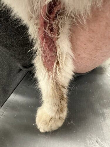
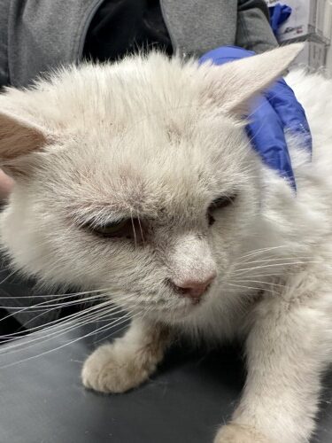
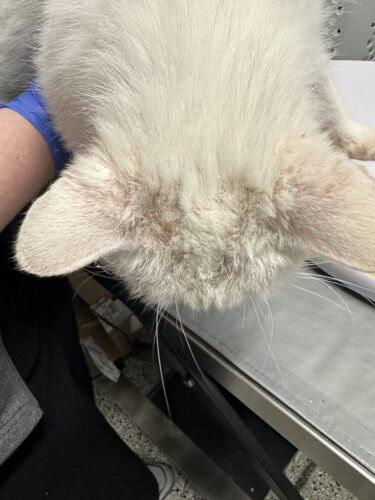
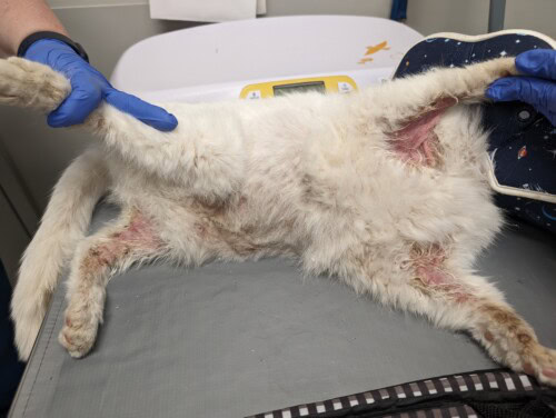
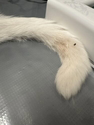
“Zelda,” a 10y FS domestic shorthaired cat, was presented to dermatology for acute-onset crusts, erythema, and pruritus. Zelda initially presented to the rDVM, on day 1, for rust-colored fur around the dewclaws.
January 2025
Shelbi Woods, BA, CVT
Finoa Lee, Dip ACVD
Dermatology Animal Clinic, Woodbridge, NJ
History
The rDVM prescribed a novel protein rabbit diet to rule out a food allergy as Zelda was already on monthly flea control (containing selamectin and sarolaner). On day 30 at the rDVM, Zelda was evaluated for nonpruritic black crusts around her mouth, eyes and nose; papules on her neck and chin; brown auricular discharge; and periocular erythema. An otic solution (containing thiabendazole, dexamethasone, and neomycin sulfate), erythromycin ophthalmic ointment, and chlorhexidine pads were dispensed. Since the owner noticed the skin changes after the transition to the novel protein rabbit diet, a hydrolyzed soy diet was prescribed instead. Zelda was eating normally, but was described by the owner to be polydipsic. General blood work (Figure 1.1) was performed because of mild weight loss and revealed increased liver values. An abdominal ultrasound was performed on day 36 (Figure 2.1), showing a pancreatic mass and hyperechoic liver. A liver supplement (containing s-adenosylmethionine (SAMe)) was recommended and started. Zelda was prescribed oclacitinib 1.3 mg/kg q12h for 14 days, then q24h thereafter; this is noted to be an off-label medication in cats.
On day 43, Zelda’s otitis resolved and chin mildly improved, but she was licking her perineum and developed periocular crusts. She was evaluated at the rDVM and a cefovecin injection was administered for the presumed perineum infection. Transdermal mirtazapine was
prescribed for decreased appetite as needed, prednisolone 0.61 mg/kg q12h for 5 days (then q24h for 5 days, then q48h for 5 days), and gabapentin 6.1 mg/kg q12h were dispensed. Oclacitinib was discontinued as the owner noted no significant improvement. On day 46, Zelda was eating better but started wheezing, so an ophthalmic solution (containing neomycin, polymyxin, dexamethasone) was prescribed on day 49, to be used off-label in both nostrils q12h for 7-10 days, and the hydrolyzed soy diet was continued. On day 55, Zelda started overgrooming and losing more hair as prednisolone was tapered to the q48h dosing; the rDVM prescribed prednisolone 0.61 mg/kg q12h for an unspecified duration (use lowest effective dose to control clinical signs).
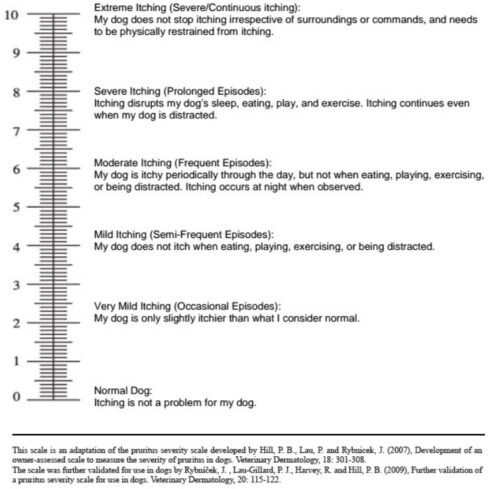
Zelda was seen on day 63 at the dermatology consult where she was losing fur on her chest, and her pruritus was considered a 10/10 on the PVA S scale (Figure 3.1), despite still being on prednisolone.
Exam
On physical examination, Zelda was quiet, alert, and responsive. She weighed 4.1 kg. She had mild referred upper respiratory noise/congestion. A grade II/VI heart murmur was auscultated for the first time.
On dermatologic examination, Zelda had severe loose white scale and hypotrichosis on her convex pinnae, dorsal head and neck, and extending down her spine. There was brown scale on the concave pinnae, around the nares, periocular, and perioral. The medial right front leg and medial left hind legs were severely moist and erythematous, alopecic, and eroded. There was yellow-brown discoloration of all four legs, likely secondary to salivary staining. There was mild erythema and hypotrichosis on her flanks, likely secondary to self- trauma.
Diagnostics
A direct impression of the medial left thigh and medial right forearm revealed cocci and severe nuclear streaming. A trichogram of the dorsal head and right forearm was negative for Demodex mites. Wood’s lamp was negative. An swab of the medial left thigh and medial right forearm was submitted for aerobic culture. A biopsy was recommended and scheduled. With the new heart murmur finding and to reduce masking changes on histopathology, the owner was instructed to taper off prednisolone. On day 66, Zelda had two affected areas on the cranial dorsum lightly shaved for histopathology submission to a dermatopathologist.
Assessment
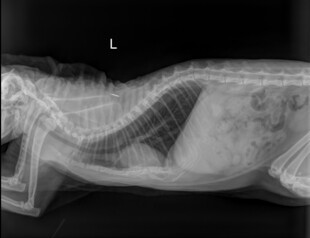
One of the leading differentials for Zelda was feline exfoliative dermatitis. The dermatologist recommended 3-view chest radiographs to be performed with the rDVM to look for a thymoma that may have triggered the skin changes. Radiographs on day 67 revealed no mass present (Figure 4.1).
Histopathology (Figure 5.1) noted mild interface dermatitis and mural folliculitis with hyperkeratosis. These changes were indicative of feline exfoliative dermatitis. With the histopathology results, and no mass seen on radiographs, Zelda was diagnosed with nonthymoma-associated feline exfoliative dermatitis.
Plan
Pending biopsy results, chlorhexidine mousse was dispensed to be used on the medial legs and axillae q12h. The owner was instructed to start chlorhexidine 2% mousse q24h and restart prednisolone 0.6 mg/ kg PO q12h for 1 day, q24h for 5 days, then q48h until histopathology
results were received. Culture results on day 70 stated methicillin- resistant Staphylococcus pseudintermedius (MRSP) (Figure 6.1). Clindamycin 9.1 mg/kg PO q12h was prescribed along with ondansetron 0.49 mg/kg PO q12h as needed for nausea. The owner was having a difficult time applying chlorhexidine mousse and keeping the cone collar on, and there was concern about being able to administer clindamycin. It was offered to hold on filling both medications pending the histopathology results, in case another more important/effective oral medication were needed to treat the underlying condition.
Follow-up
On day 73, histopathology results were received and relayed to the owners who noted Zelda was not tolerating the cone collar but allowed for the application of the chlorhexidine mousse. Zelda was still eating but not as much as previous, so transdermal mirtazapine was restarted. On day 78, Zelda was still pruritic and the owners noted she was able to reach around the cone collar. Prednisolone was increased to 1.2 mg/kg PO q12h. The clindamycin and ondansetron previously prescribed were instructed to be filled. At the derm recheck exam on day 80, the owners noticed improvement of Zelda’s crusts with the first two days on antibiotics, but they felt that she regressed ever since, also noting lethargy and hiding. Physical exam revealed ~30 – 40% improvement of the brown scale/ debris periocular, perilabial, on concave pinnae, and around the nares. Brand name cyclosporine 7.3 mg/kg PO q24h was started. Prednisolone, clindamycin, and mirtazapine were continued as previously prescribed.
Zelda was seen for a second derm recheck exam on day 105. Her owners reported her ventrum was improving after the recheck on day 80, but Zelda had an acute increase in pruritus and erythema just 3 days prior, on day 102. Chlorhexidine solution 2% q12h was dispensed to
be sprayed or wiped on Zelda’s skin, in place of the chlorhexidine mousse. On day 108, Prednisolone was increased to 1.8 mg/kg PO q24h ongoing. The owners were instructed to continue brand name cyclosporine as previously prescribed. Due to quality-of-life concerns, Zelda was humanely euthanized at the rDVM on day 114.
Discussion
Exfoliative dermatitis isn’t a disease but rather a cutaneous reaction pattern. It can either be triggered by an underlying cause, or idiopathic. Some triggers of this reaction pattern include a thymoma, epitheliotropic lymphoma, visceral malignant neoplasms or drug reactions.1 There are no breed or sex predilections and has been reported to affect cats as young as 4-years-old but typically affects middle-aged to older felines.1 Exfoliative dermatitis is rare in cats, but thymomas are one of the most common mediastinal neoplasms in cats. Exfoliative dermatitis has also been reported in rabbits. It can be associated with malnutrition, dermatophytosis, ectoparasites, Malassezia dermatitis, sebaceous adenitis, epitheliotropic lymphoma, and thymomas, but thymomas in rabbits are uncommon.3
Clinical characterization of exfoliative dermatitis is severe exfoliation, scale, and erythema, which can either be localized or generalized. A study revealed pruritus was present in 50% of non-thymoma associated cases in felines.4 Lethargy, anorexia, and weight loss for several weeks has previously been reported.4
There are two reported types of exfoliative dermatitis: thymoma-associated and nonthymoma-associated. Thymoma-associated is confirmed by the identification of a mass of the thymus gland via imaging.5 If a tumor is present, a CT scan is the preferred method for
staging the tumor because it can assess tumor invasiveness and help with surgical planning.7
Other diagnostics to rule out differential diagnoses include skin cytology and scrapings, dermatophyte culture, trichogram, and serology for feline immunodeficiency virus (FIV) and feline leukemia virus (FeLV).5
The pathomechanism by which a thymoma causes cutaneous lesions is unclear, but there is suspicion that the diseased thymus activates autoreactive cytotoxic T cells that act on epithelial cells.4 The most common histological changes include orthokeratotic and parakeratotic hyperkeratosis, epidermal acanthosis, variable psoriasiform epidermal hyperplasia, variable lymphocytic exocytosis, and perivascular to lichenoid dermal inflammation.1 The epidermal basal layer contained a hydropic degeneration of keratinocytes.5 Infundibular lymphocytic mural folliculitis and decreased or absent sebaceous glands can be seen.6 Interface dermatitis composed of lymphocytes and varying amounts of mast cells and plasma cells, is seen in the dermis.6
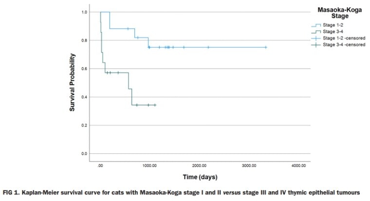
In thymoma-associated exfoliative dermatitis, surgical removal of the tumor can result in improvement and potential resolution. The tumor is recommended to be removed via lateral thoracotomy, opposed to median sternotomy, which is associated with minimal complication rates and long-term post-operative pain.5 In one study, the human Masaoka-Koga staging system was used; it associated the outcome for cats undergoing thymoma surgery. In this same study, perioperative mortality ranged from 11%-22% and was typically seen in cats with a Masaoka-Koga stage III to IV invasive thymoma (Figure 7.1).7 For incompletely resected or non-resectable tumors, radiation therapy can be considered.7
Immunosuppressive therapy is the main treatment of nonthymoma-associated exfoliative dermatitis. Prognosis is variable as it depends on the severity of the dermatitis and response to treatment. A study of nonthymoma-associated exfoliative dermatitis cats responded to: 1. Immunosuppressive doses of prednisolone (4 mg/kg/day) only, 2. A combination of prednisolone and cyclosporine, 3. cyclosporine only (7 mg/kg/day), or 4. oral dexamethasone (0.2 – 0.5 mg/kg).4,8
Figure 1.1
Hematology results from Antech Diagnostics (East)
3760 Requisition ID: 19250
                                        Posted      Final
Test                          Result            Reference Range
MCHC                          32 g/dL           30 - 38
WBC                           4.2 10^3/uL       3.5 - 16.0
LYMPHS                        21 %              20 - 45
MONOS                         4 %               1  -  4
NEUT                          70 %              35 - 75
EOS                           5 %               2  - 12
BASO                          0 %               0  -  1
RBC                           9.7 10^6/uL       5.92 - 9.93
MCV                           49 fL             37 - 61
MCH                           15.8 pg           11 - 21
BLOOD PARA                    None Seen
NEUT BANDS                    0 %               0 - 3
PLATELETS                     137 10^3/uL L     200 - 500
PLATE (EST)                   Adequate
ABS BASO                      0 /uL             0  - 150
ABS EOS                       210 /uL           0 - 1000

ABS LYMPHS                    882 /uL L         1200 - 8000
ABS MONOS                     168 /uL           0  - 600
ABS NEUTS                     2940 /uL          2500 - 8500
HEMOGLOBIN                    15.3 g/dL         9.3  - 15.9
HEMATOCRIT                    48 %              29 - 48
Figure 2.1
Chemistry results from Antech Diagnostics (East)
3760 Requisition ID: 19250                      Posted    Final
Test                      Result                Reference Range
ALB                       3.8 g/dL              2.5 - 3.9
ALKP                      256 IU/L H            6 - 102
ALT                       755 IU/L H            10 - 100
AMYL                      676 IU/L              100 - 1200
AST                       254 IU/L H            10 - 100
BUN/UREA                  21 mg/dL              14 - 36
Ca                        9.7 mg/dL             8.2 - 10.8
Chloride                  117 mEq/L             104 - 128
CHOL                      143 mg/dL             75 - 220
CK                        170 IU/L              56 - 529
CREA                      1.1 mg/dL             0.6 - 2.4
GGT                       2 IU/L                1 - 10
GLU                       76 mg/dL              64 - 170
Mg                        2.0 mEq/L             1.5 - 2.5
PHOS                      5.1 mg/dL             2.4 - 8.2
Potassium                 5.4 mEq/L             3.4 - 5.6
Sodium                    152 mEq/L             145 - 158
TBIL                      1.0 mg/dL H           0.1 - 0.4
TP                        8.4 g/dL              5.2 - 8.8
TRIG                      48 mg/dL              25 - 160
GLOB                      4.6 g/dL              2.3 - 5.3
A/G Ratio                 0.8                   0.35 - 1.5
B/C Ratio                 19                    4 - 33
Na/K Ratio                28  L                 32 - 41
Findings
Liver: Moderately large and isoechoic to the spleen; Gall bladder: There is a moderate volume of anechoic bile within the lumen; Common bile duct: Unremarkable/Not visualized
Spleen: Unremarkable
Left Kidney: Measures 3.6cm in length; Unremarkable
Right Kidney: Measures 4.0cm in length; Appearance is similar to left kidney
Left Adrenal: Unremarkable, measures 0.4cm in thickness
Right Adrenal: Unremarkable, measures 0.3cm in thickness
Urinary Bladder: Unremarkable
Stomach: Measures normal in thickness and normal layering is preserved; mild fluid and gas within the lumen
Small Intestines: Measure normal in thickness and normal layering distinction appears preserved; Minimal, normal volume of fluid present within the lumen; Peristalsis subjectively normal
Colon: Measure normal in thickness and normal layering appears preserved; gas and stool in colon
Pancreas: There is a moderately well-defined, 0.55 x 0.65cm hypoechoic nodule in the distal left limb of the pancreas
Sublumbar and iliac lymph nodes: Unremarkable
Mesenteric and abdominal lymph nodes: Moderately enlarged and hypoechoic mesenteric lymph node measures 0.35 x 0.8cm
Abdominal cavity: No evidence of peritoneal fluid is seen
Summary/Opinion: Diffuse hepatopathy with hyperechoic liver – r/o steroid, diabetic, lipid or non-specific vacuolar hepatopathy vs infiltrative disease process (lymphoma, etc) vs cholangiohepatitis vs other. Prominent mesenteric lymph node – r/o reactive vs metastatic vs lymphoma vs other. Pancreatic nodule in distal left limb – r/o nodular hyperplasia vs adenoma vs pancreatic carcinoma vs lymphoma vs other.
Figure 3.1
Figure 4.1
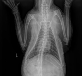
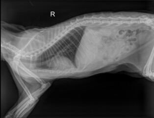
Figure 5.1
Tissue and site
Interscapular

Sample type
Punch

Diagnosis
MILD INTERFACE DERMATITIS AND MURAL FOLLICULITIS WITH HYPERKERATOSIS - SCAPULAR SKIN - FELINE.

Two skin punch biopsies of excellent quality are examined. The samples have diffuse mild epidermal hyperplasia with
orthokeratotic to parakeratotic hyperkeratosis. The epidermis has lymphocyte exocytosis, occasional Langerhans cell
aggregates, and few apoptotic keratinocytes. Many hair follicles retracted into mid dermis, and scattered hair follicle
atrophied. Anagen bulbs persist. Some outer root sheaths have lymphocyte exocytosis and loss of crisp dermal
distinction. The superficial dermis has a mild infiltrate of lymphocytes, plasma cells and mast cells. The basal lamina is
multifocally thickened. The samples do not have sebaceous gland loss.

The histologic features fall into the category of feline exfoliative dermatosis. There is no evidence of lymphoma.
Figure 6.1
Status: FINAL

Isolate 1: Staphylococcus pseudintermedius - 3+

                                        Isolate 1   MIC
Penicillin                              R           >=0.5
Oxacillin / Methicillin                  R           >=4
Cefovecin                               R           >=8
Amikacin                                S           <=2
Gentamicin                              I           8
Enrofloxacin                            R           >=4
Marbofloxacin                           R           >=4
Erythromycin                            S           0.5
Clindamycin                             S           0.25
Doxycycline                             R           2
Mupirocin                               S           <=1
Chloramphenicol                         S           8
Florfenicol                             S           <=4
Rifampin                                S           <=0.03
Trimethoprim/Sulfamethoxazole           R           >=320
Amoxicillin                             R          >0.25
Amoxicillin-Clavulanic Acid             R          >0.25
Imipenem / Carbapenem
Cephalexin                             R            >2
Cefpodoxime                              R           >=8
Ciprofloxacin                           R
Azithromycin                      		 S
Minocycline                             R
Figure 7.1
References
- Whitbread T. Jubb, Kennedy and Palmer’s Pathology of Domestic Animals. Veterinary Dermatology. 2010;1:553. doi:10.1111/j.1365-3164.2009.00767.x
- Miller WH, Griffin CE, Campbell KL. Muller and Kirk’s small animal dermatology. Elsevier Health Sciences; 2012.
- Florizoone K. Thymoma-associated exfoliative dermatitis in a rabbit. Veterinary Dermatology. 2005;16(4):281-284. doi:10.1111/j.1365-3164.2005.00456.x
- Linek M, Rüfenacht S, Brachelente C, et al. Nonthymoma-associated exfoliative dermatitis in 18 cats. Veterinary Dermatology. 2014;26(1):40. doi:10.1111/vde.12169
- Cavalcanti JVJ, Moura MP, Monteiro FO. Thymoma associated with exfoliative dermatitis in a cat. Journal of Feline Medicine and Surgery. 2014;16(12):1020-1023. doi:10.1177/1098612×14531762
- Rottenberg S, Von Tscharner C, Roosje PJ. Thymoma-associated exfoliative dermatitis in cats. Veterinary Pathology. 2004;41(4):429-433. doi:10.1354/vp.41-4-429
- Marks TA, M Rossanese, Yale AD, et al. Prognostic factors and outcome in cats with thymic epithelial tumours: 64 cases (1999-2021). The journal of small animal practice/Journal of small animal practice. 2023;65(1):47-55. doi:https://doi.org/10.1111/jsap.13675
- 1.Szczepanik M, Piotr Wilkołek, Śmiech A, Kalisz G. Non-thymoma-associated exfoliative dermatitis in a European shorthair cat: A case report. Veterinary Medicine and Science. 2021;7(6):2108-2112. doi:https://doi.org/10.1002/vms3.583


