An 11-year 5-month-old male neutered domestic long-haired cat presented with a five-year history of generalized pruritus and hard palate ulceration.
Meghan Solc, DVM, DACVD
Dermatology for Animals, Catonsville, Maryland
November 2024
History
-
An 11-year 5-month-old male neutered domestic long-haired cat presented with a five-year history of generalized pruritus and hard palate ulceration. The generalized pruritus had been historically controlled with a hydrolyzed diet and off-label use of oclacitinib.
-
The patient presented to the dermatology practice on oclacitinib 0.9mg/kg twice daily, hydrolyzed soy-based diet but no current parasite prevention. His left eye had been enucleated due to a diffuse iris melanoma. No skin or oral mucosal histopathology had been performed.
-
The pet parent reported that the patient’s pruritus recently became intolerable. The patient required an Elizabethan collar to prevent self-trauma.
-
Despite therapies, the previously noted oral ulceration had progressed.
Exam
General Exam:
- Temperature: 101.6 degrees Fahrenheit
- Respiration: 40 breaths per min
- Pulse: 230 beats per min
- Mucous membranes: pale pink and moist
- Body Condition 4/9
- Capillary refill time: less than 2 sec
- Weight: 4.04 kg
- Thoracic evaluation: clear lung sounds and a grade III/VI heart murmur
- Enucleated OS
Dermatologic Exam:
- Shiny, long well-kept black hair coat
- Ear canals were pliable and patent AU with tympanic membranes visualized.
- Interdigital plantar erythema and black exudate matted to the hair with no ulcerations.
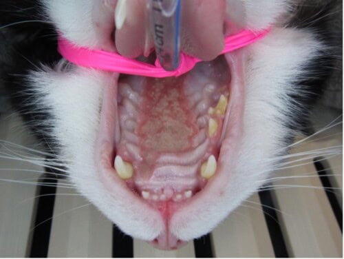
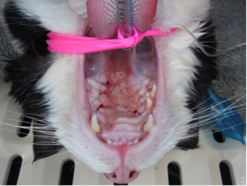
- Hard palate lesions extend from the level of the maxillary canines distal to the soft palate and to mucogingival border.
- No lingual masses were noted.
Figures 1 and 2: Initial presentation prior to biopsy
Differential Diagnoses
- Eosinophilic granuloma vs. Menrath’s ulcer vs. Neoplasia
- Allergic dermatitis
Diagnostics
The location of the lesion raised concerns for palatine arterial bleeding upon biopsying. It was decided that the patient would be referred to a board-certified veterinary dentist for biopsies. Tissue samples for histopathology and culture and a more thorough oral examination were obtained.
- Histopathology: Submucosa contained high numbers of eosinophils, fewer macrophages, plasma cells, lymphocytes, and neutrophils. Rare collagen flame figures. The surface was extensively ulcerated.
- Anaerobic bacterial culture: Bacteroides species
- Aerobic bacterial culture: Enterococcus species
Assessment
- Menrath’s ulcer with bacterial infection
- Allergic dermatitis
Treatment Plan
- Dexamethasone orally 0.2mg/kg at a tapering schedule over a 4–6-week period
- Modified cyclosporine introduced gradually due to the patient’s history of gastrointestinal upset and dose of 7.5mg/kg daily was maintenance dose goal.
- Oclacitinib was discontinued while on other immunomodulatory therapies.
- Cefovecin subcutaneously every 2 weeks for 3 treatments
Follow-up
At the one-month follow-up appointment the patient’s oral ulcer had diminished in size by at least 65 %. The initial improvement that was noted was attributed to the systemic steroids and antibiotics. The cyclosporine was started to provide long-term control however it was not well tolerated and was discontinued. The oclacitinib treatment was restarted in hopes of preventing a full relapse.
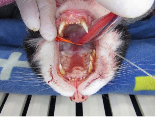
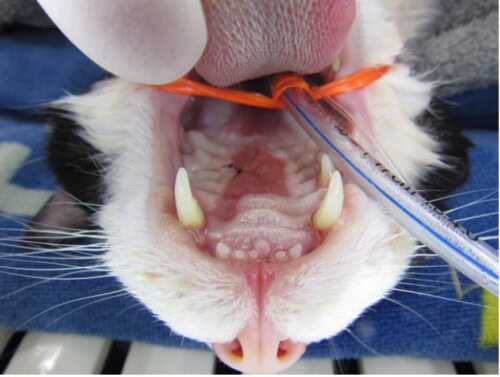
One month after the dermatology follow-up, the patient presented to the dental clinic for acute bleeding from the mouth. The patient survived an emergency palatal artery ligation but relapsed quickly within 24 hours of this procedure. He was referred to an E.R. facility for acute care. The suture that was placed to ligate the artery was no longer present.
Figure 3: Acute palatine arterial hemorrhage presentation
Figure 4: Post left palatal artery ligation.
Figure 5: Lateral view of eosinophilic ulceration with severe defect
At the E.R, an esophageal feeding tube was placed to prevent further oral trauma. A whole blood transfusion was administered due to the persistent decline in the PCV (15%). The patient remained in hospital for esophageal feeding tube management, subsequent blood transfusions, and sedation for 48 hours. The cyclosporine was restarted in hopes it would help control the oral ulceration.
Ultimately the patient was discharged to pursue home care. The patient was sent home to be managed palliatively with esophageal feedings, gabapentin, Yunnan Baiyao, and cyclosporine modified. The patient remained stable with a PCV of 28% for 48 hours after discharge. Within 72 hours of discharge the patient started to hemorrhage and ultimately declined, passing away 7 days following his E.R. hospitalization.
Retrospectively this lesion had been present in this patient for approximately four years.
Discussion
-
Menrath’s ulcer has been coined to describe a lesion on the hard palate that is associated with detrimental hemorrhage of the palatine artery. Eosinophilic granulomas and oral ulcerations are commonly noted lesions on the hard palate of allergic cats but rarely result in acute hemorrhage and death.
-
Overgrooming and eosinophilic granulomas are common primary symptoms of allergic feline patients. The symptom of overgrooming may result in self trauma and secondary lesions. Eosinophilic hard palatal granulomas may be exacerbated by the secondary trauma from the papilla of the tongue and overgrooming. Secondary trauma to the hard palate ulcer can lead to overt hemorrhage. Hemorrhage may not be noted by the pet parent or the veterinarian due the location of the bleed in the oral cavity. The digestion of blood may result in discrete symptoms and may not be as obvious to detect. The diagnosis of Menrath’s ulcer may be complicated by the lack of outward symptoms and difficulty monitoring for active hemorrhage.
-
Surgical intervention and ligation are required to help manage active hemorrhage associated to the palatine artery. If ligation is not successful, the prognosis for these cases is extremely guarded. Managing and treating the primary symptoms should be the continued focus for treatment of the allergy and primary symptoms. Aggressive treatment of allergic disease with dietary modification, parasite treatment, immunotherapy and/or antiallergy therapies should be pursued.
Key Takeaways:
- Hard palate ulceration is a clinical feature associated with feline allergic symptoms.
- Recommend more aggressive treatment for patients with hard palate ulceration to prevent potential complication of hemorrhage.
- Symptomatic management of inflammation and allergy should be pursued at all costs while investigation of the allergic disease is investigated.
- Prioritize prevention of future relapses with more aggressive measures.
References
-
Menrath, V.H., Miller, R. The repair and prevention of bleeding palatine erosive lesions in the cat. Australian Veterinary Practitioner. 1995, Vol 25, No 4: 202-204, 206 ref.1.


