“Gertrude” is a 5-year-old FS mixed breed dog who presented for a refractory, progressive alopecia and crusting on the face, trunk, and limbs for about 1 year prior to presentation.
Carine Laporte, VMD, DACVD
Dermatology for Animals, Salt Lake City
October 2024
History
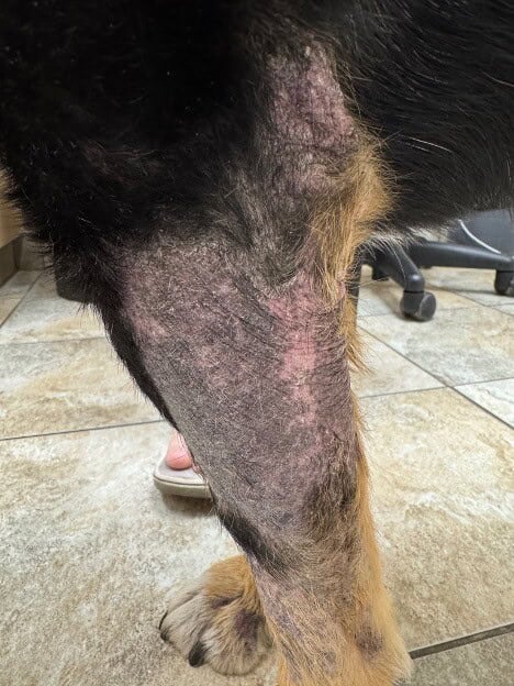
- Gertrude developed progressive alopecia, crusting, hyperpigmentation, and pruritus on the head, trunk, and libms approximately 1 year prior to presentation to the veterinary dermatologist.
- There had been insufficient response to previous oral glucocorticoids (anti-inflammatory doses); beta-lactam antibiotics (numerous courses); strict 12-week, Purina HA elimination diet trial; Apoquel 0.6mg/kg PO attempted at both q24 hour and q12 hour dosing intervals; a Simparica ectoparasite treatment trial; and a three-week course of fluconazole 5 mg/kg/day.
- There were no systemic signs.
- Apart from her pruritus, Gertrude did not seem bothered by her skin.
- Complete blood count, chemistry panel, total T4, and urinalysis were normal 1 week prior to presentation.
- Gertrude had no previous dermatologic history.
- She was housed indoors but spent a significant amount of time free ranging outdoors. She therefore had potential extensive exposure to wildlife and was regularly found ingesting carcasses of various wild animals.
- Due to the duration of the lesions and progression despite attempted therapies, the owner was considering euthanasia if a diagnosis could not be obtained.
Dermatologic Exam
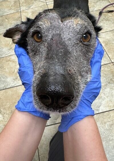
On physical examination, there were multifocal to coalescing patches of alopecia, erythema, hyperpigmentation, scale, and yellow-grey crusting, with occasional erosions and ulcerations. The head and face were most affected; discrete lesions were also appreciated on the trunk and limbs. All claws had varying degrees of onychogryphosis.
Vital signs and body condition scoring were within normal limits.
Differential Diagnoses
Differential diagnoses included dermatophytosis, demodicosis, ischemic dermatopathy, vasculitis, and pemphigus foliaceus., +/- secondary superficial pyoderma.
Diagnostics
- Wood’s Lamp: negative.
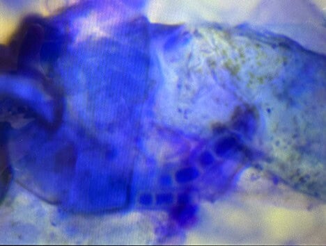
- Tape prep cytology: pyogranulomatous inflammation with fungal hyphae.
- Deep skin scraping: no mites.
- Aerobic bacterial culture: no growth.
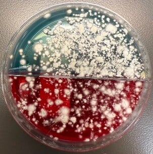
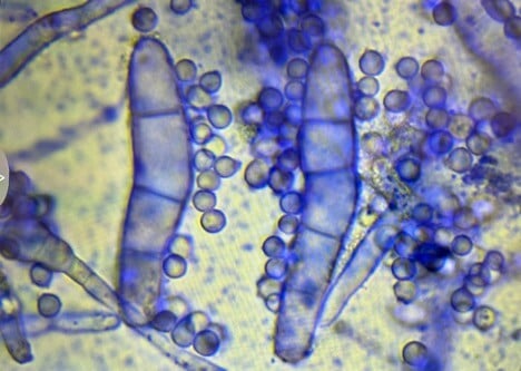
- Dermatophyte culture (DTM) and PCR: positive for Trichophyton mentagrophytes complex.
Figure 3. Fungal hyphae on cytology.
Figure 4. Trichophyton mentagrophytes complex on a dermatophyte culture plate (DTM). Note color change of the media immediately with colony growth.
Figure 5. Trichophyton mentagrophytes complex macroconidia and microconidia on a sample taken from a colony grown on the DTM. Image is from microscopic evaluation at 100x.
Assessment
Dermatophytosis
Treatment Plan
The treatment plan consisted of four aims:
- Treat the Patient: Terbinafine 40 mg/kg PO SID, weekly baths with miconazole nitrate 2% /chlorhexidine gluconate 2% shampoo, and daily mousse containing the same miconazole/chlorhexidine ingredients. Treatment to continue until two negative consecutive cultures taken 2-4 weeks apart.
- Test In-Contact Animals: The owner was advised to have other animals tested by their local veterinarian using both DTM and PCR.
- Environmental Decontamination: Twice weekly vacuuming, laundering, and disinfecting with accelerated hydrogen peroxide; discarding any non-porous products that could not be adequately cleaned (e.g., cat tree).
- Prevent Reinfection: Outdoor lifestyle restrictions were suggested but would be challenging due to owner preference.
Follow-up
Gertrude has responded well to treatment after two months, with lesions progressively improving. Treatment is ongoing, as is monitoring through q2 week DTM, PCR, and clinical evaluation.
Discussion
Dermatophytosis, or ringworm, is a zoonotic fungal infection commonly caused by Microsporum canis, Microsporum gypseum, or Trichophyton mentagrophytes. Infection occurs via direct contact or fomite transmission, but not all exposures lead to infection. Risk factors include young age, free-roaming behavior, immunosuppression, and skin trauma.
While clinical signs of dermatophytosis in dogs may include the typical “ring-shaped” lesion described in people, lesions also can have a markedly variable appearance: erythema, alopecia, scale, crust, hyperpigmentation, comedones, changes in claw appearance, nodules, and sometimes pruritus. Dermatophytosis does not cause systemic disease, though it can be associated with immunosuppressive diseases which can themselves be associated with systemic signs.
There is no single gold standard test for diagnosis of dermatophytosis. Diagnosis is best obtained using a combination of test modalities: cytology, Wood’s lamp examination, DTM culture, PCR, dermoscopy, and/or histopathology.
Importantly, Gertrude’s case exemplifies that cytology can offer immediate results. Additionally, Gertrude’s case illustrates that a negative Wood’s Lamp examination does not definitively rule out dermatophytosis; positive fluorescence is primarily seen with strains of M. canis dermatophytes but will not be seen, for example, with T. mentagrophytes complex as Gertrude had.
There also is no gold standard test for monitoring response to therapy. Monitoring usually involves a combination tests: clinical response, DTM, PCR, and possibly Wood’s Lamp in cases of M. canis dermatophytosis that initially fluoresced prior to treatment. DTM is particularly useful for monitoring response to therapy because a reduction in the number of colony forming units can point towards good response to treatment even though the culture remains “positive.” In contrast, a positive PCR result may not be a sufficiently informative way to monitor response to treatment as it could equally indicate true ongoing infection, contamination, or non-viable (dead) hyphae. However, a negative PCR result in a treated animal with clinical resolution of lesions would be compatible with cure
Treatment of dermatophytosis requires systemic and topical antifungal therapies, environmental decontamination, and precautions to prevent spread. Some important considerations regarding treatment are:
- Systemic treatment with terbinafine or itraconazole is effective; ketoconazole and fluconazole may be less effective treatment options with a broader side effect profile (ketoconazole).
- Topical treatment with miconazole/chlorhexidine shampoos, lime sulfur, or enilconazole applied twice weekly enhances therapeutic outcomes.
- Environmental cleaning primarily aims to reduce environmental contamination rather than prevent direct infection from the environment. Infections from the environment alone are likely quite rare. In contrast, false positive DTM or PCR test results from contaminant fungi on the hair coat obtained through environmental contamination are common and will result in prolonged treatment durations.
- Dogs and cats will almost always have clinical lesion resolution prior to mycologic cure. Because of this, treatment duration should not just be based on clinical appearance. Treatment is continued until 2-3 negative, consecutive cultures or PCRs taken 1-4 weeks apart.
- Environmental decontamination can be accomplished by clipping the hair around the patient’s affected lesions, topical treatment of the patient, and cleaning of the environment. Antifungal disinfectants include sodium hypochlorite (household bleach) in 1:10 to 1:100 dilution, enilconazole 20 uL/L, and accelerated hydrogen peroxide. Disinfection of nonporous substances involves twice weekly mechanical removal of all debris via vacuum or sweeping, washing of the target surface with a detergent until the area is visibly clean, then application of a disinfectant to kill residual spores. Disinfection of laundry can be achieved by two washings on the longest wash cycle and using the clothes dryer. Disinfection of carpets can be done through hot water extraction after vacuuming to remove gross debris.
- Asymptomatically infected or asymptomatic carriers are possible with this disease. As such, all in-contact dogs and cats should be tested for dermatophytosis.
- Confinement of infected animals is an important part of containing outbreaks of dermatophytosis. Items in the confinement area should be limited to those that can be washed daily (e.g. towel, blanket) and all toys should be plastic. However, there may be negative behavioral repercussions of isolating an animal for extended periods of time, particularly in puppies and kittens. Owners and veterinarians should work together to develop a protocol for deliberate social exposure.
Key Takeaways: While dermatophytosis is relatively common, Gertrude’s case is interesting because it illustrates four important considerations about the disease:
1. Clinical presentations: Dermatophytosis can have variable clinical presentations, in some cases mimicking serious autoimmune conditions like pemphigus foliaceus, vasculitis, and ischemic dermatopathy. In certain patients, lesions may even resemble those of cutaneous epitheliotropic T-cell lymphoma. You can compare this case to a previous case of Trichophyton dermatophytosis we posted in January 2024 to see some of the diversity in clinical presentations. However, when a patient presents with alopecia and crusting of haired skin, ruling out the main causes of folliculitis is a good first step. The three main causes of folliculitis are pyoderma, demodicosis, and dermatophytosis.
2. Diagnosis: Cytology is a fundamental initial diagnostic test. Identification on cytology ultimately allows for more rapid treatment initiation, which helps not only the individual patient but also helps to reduce risk of spread to other animals and humans, and reduce environmental decontamination.
3. Therapeutic Response: While a positive response to an antifungal therapy can support the diagnosis of dermatophytosis, lack of response does not definitively rule it out. Dermatophytosis requires prolonged anti-fungal treatment, and not all patients will respond to a particular antifungal drug. Gertrude’s apparent lack of response to fluconazole prior to presentation to the dermatologist may have been because fluconazole is potentially a lesser effective treatment option for dermatophytosis, and/or because of the short duration of treatment (3 weeks).
4. Environment: Preventing reinfection requires diligent environmental management and limiting exposure to infected animals or wildlife.
References
- Moriello KA et al. Diagnosis and treatment of dermatophytosis in dogs and cats. Clinical Consensus Guidelines of the World Association for Veterinary Dermatology. Vet Dermaol 2017; 28: 266-e68.
