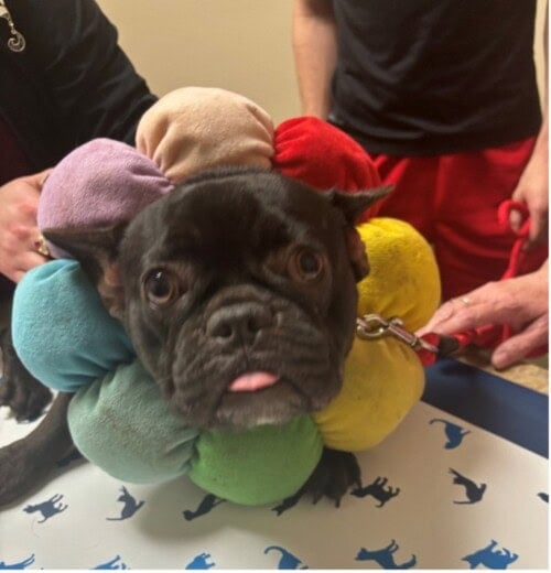
A 1.5-year-old female spayed French bulldog was presented for chronic otitis with severely swollen and stenotic ear canals.
Curtis Plowgian, DVM, DACVD
Animal Dermatology Clinic Indianapolis
September 2024
History
- 1.5-year-old female spayed French bulldog
- Problems with the ears began around the time of spaying (~6 months of age).
- At the time of the spay surgery, Lilo was treated with topical ear medications from her primary veterinarian which seemed initially to help.
- Lilo’s ears currently bleed and she spreads the blood around the house by shaking and rubbing her ears.
- Owner has 3 Great Danes and 2 other mixed breed dogs, none of which are affected.
- All dogs in the household are on or starting on Simparica Trio.
- Owner has tried Benadryl, and different sprays and several medicated drops for the ears (Zymox most recently).
- Current diet is an over-the-counter kibble (owner is unsure of brand).
- Owner is unsure of number or quality of bowel movements per day (dogs go free-choice in the yard, and owner pays someone to scoop the yard) but is not aware of any diarrhea.
- Lilo has frequent malodorous gas at home.
- No current medications and no health conditions aside from the ears.
Dermatologic Exam
-
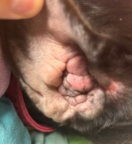
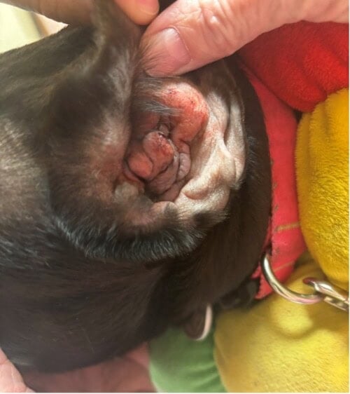
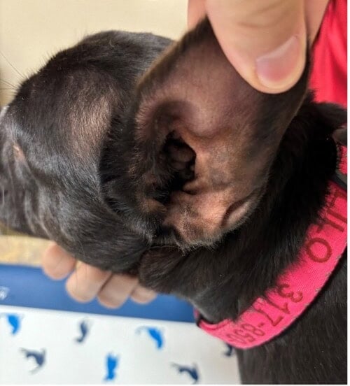
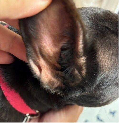
Both ear canals are firm and swollen on palpation, and ears are completely occluded via stenosis and proliferative swollen tissue (no visible ostia to the canals and a swab can barely be passed with enough prodding).
-
No induration or mineralization noted on palpation of ear canals.
-
Skin and coat look ok.
Diagnostics
Cytology Findings:
-
Ear swab: too numerous to count coccoid-shaped bacteria and rod-shaped bacteria with 0-5 yeast per oil immersion field (OIF) AU
Assessment
- Severe chronic stenosis with end-stage inflammatory changes
- Suspect underlying allergies, rule out food allergies vs environmental allergies vs combination.
Treatment Plan
It was discussed with owners that with the degree if inflammation and stenosis seen on both ear canals it may not be possible to salvage the ear canals, and that Lilo might need a bilateral total ear canal ablation and bulla osteotomy (TECABO). However, the dermatologist would try their best to reduce stenosis with anti-inflammatory therapies in the form of a hypoallergenic elimination diet trial (Purina HA), aggressive oral steroids (0.95mg/kg prednisone SID x 6 weeks), and aggressive topical steroids combined with antimicrobials in the form of ear drops (enrofloxacin/miconazole/fluocinolone acetonide and DMSO drops, 0.3mL AU q12 hours). Twice weekly cleanings with TrizUltra+Keto Cleaner was also recommended for debris removal. The owner was advised to inform the dermatologist if the Lilo had any side effects to the oral or topical steroids, and to call if there were any problems with the diet, trial, or if the ears were worsening at any point.
Follow-up
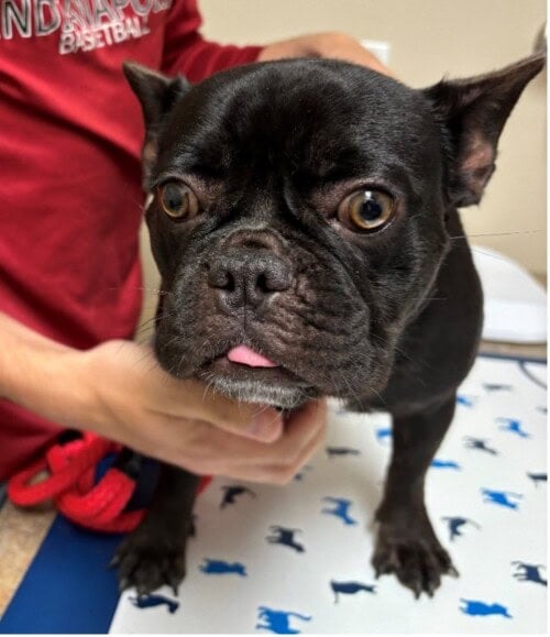
At 6-week recheck, Lilo’s ears were markedly improved; the owner didn’t report any serious adverse effects of any of the medications that had been started, and the ears had opened to the point that they were easy to clean and no longer bleeding or producing significant amounts of exudate. Lilo was eating the Purina HA well with no known deviation from the strict diet trial, and bouts of gas at home had improved/nearly resolved.
On repeat cytology, the left ear was completely free of infection, and the right ear had 0-5 yeast per OIF. Surolan (prednisolone acetate, polymyxin B and miconazole) was started q12 hours in the right ear, and mometasone 0.1% solution was started twice weekly as an anti-inflammatory maintenance drop in the right ear. Ear cleanings were maintained at a twice weekly cadence, and the oral prednisone was tapered off over the subsequent 2 weeks. A 6-week follow up appointment was scheduled to challenge the diet and to ensure that progress in the ears was maintained.
Discussion
While this case wasn’t particularly unique or challenging diagnostically, it was unusual for several reasons, and valuable to clinicians to consider when treating a particularly difficult case of otitis externa. Firstly, while we typically think of end-stage anatomic changes to the ear canals as a chronic change in middle-aged or older dogs, this case happened to present in a younger (1.5y) dog, although the dog had been dealing with chronic/recurrent infections for almost a year. Perpetuating factors that develop in cases of chronic otitis externa include altered epithelial migration, and thickening of the skin and cartilages of the ear canal, eventually leading to stenosis of the lumen of the ear canal. Stenosis and inflammation of the ear canal can also lead to fold dermatitis, which makes cleaning and medicating ears more difficult, and predisposes the ear to future infections1. Further end-stage changes include mineralization of the cartilages of the ear canal, which fortunately were not seen in this case.
The second unusual aspect of this case is that often ears that have become as swollen and stenotic as Lilo’s often require TECA-BO. While Lilo didn’t have any irreversible changes to her ear canals such as mineralization, it is not always a guarantee that significant degrees of stenosis and swelling such as those seen in this case will reverse with medical treatment. In the author’s opinion, steroids are far and away the best medication to treat the types of chronic inflammatory changes discussed above, better than any other allergy or anti-inflammatory medication. In a review article of human otitis externa, while topical steroids are considered a first line treatment for the inflammation caused by otitis externa, oral or systemic steroids can be considered in refractory cases2. In this case, aggressive courses of oral and topical steroids were pursued concurrently, which is usually the treatment course preferred by the author when the ear canals have progressed to the point where surgery will be indicated if medical management fails.
In an ideal clinical scenario, Lilo’s ears would have been rechecked in 2 or 4 weeks to make adjustments as needed to the treatment plan, and especially to start tapering the oral prednisolone sooner once the ear canals opened up, but as is common in post-pandemic specialty medicine, a 6-week recheck was the soonest available appointment, and given the degree of swelling in both ears, the author felt comfortable with a slightly longer course of oral steroids than would usually be given without tapering.
While food allergies weren’t definitively diagnosed in this case yet (diet challenge still pending as of this writing), food allergies were suspected in this case given the early age of onset and the additional gastrointestinal signs (frequent gas noted at home). While environmental atopic dermatitis usually occurs in young adults (typically aged 1-3), food allergies can start at any age, and ears are a very commonly affected area of the body3. The fact that flatulence has improved at home on the diet trial is supportive evidence of an underlying food allergy, but a challenge of the diet would be needed at following appointment (after 8-12 weeks of strict adherence to the diet) to confirm for sure if food allergies are present4. Diagnosing and treating underlying food allergies can be very helpful in managing cases of chronic and refractory otitis, and should not be overlooked in a difficult ear case.
While the presentation and therapeutic response in this case were dramatic and unusual, this case illustrates that some cases of chronic and refractory otitis that might seem to be destined for surgery can be salvaged with aggressive and appropriate medical management.
References
- Miller WH, Griffin CE, Campbell KL. Muller and Kirk’s Small Animal Dermatology. 7thPhiladelphia, PA: Saunders, 2013; 749-750.
- Wiegand S, Berner R, Schneider A, Lundershausen E, Dietz A. Otitis Externa. Dtsch Arztebl Int. 2019 Mar 29;116(13):224-234. doi: 10.3238/arztebl.2019.0224. PMID: 31064650; PMCID: PMC6522672.
- Olivry, T., Mueller, R.S. Critically appraised topic on adverse food reactions of companion animals (7): signalment and cutaneous manifestations of dogs and cats with adverse food reactions. BMC Vet Res15, 140 (2019). https://doi.org/10.1186/s12917-019-1880-2
- Olivry, T., Mueller, R.S. & Prélaud, P. Critically appraised topic on adverse food reactions of companion animals (1): duration of elimination diets. BMC Vet Res 11, 225 (2015). https://doi.org/10.1186/s12917-015-0541-3.