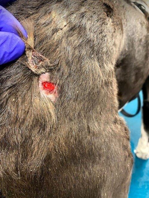
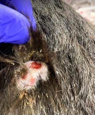
An 8-year-old male neutered border collie mix was presented for acutely rupturing cyst-like lesions focused mostly on the perianal region.
Andrew Simpson, DVM, MS, DACVD
VCA Aurora Animal Hospital
August 2024
History
- 8 year-old male neutered border collie mix
- Chronic history of seasonal atopic dermatitis for many years
- History of hypothyroidism (well-controlled with levothyroxine)
- Acutely developed rupturing cyst-like skin lesions focused mostly on the perianal region
- The cyst-like lesions were sampled for bacterial culture – no growth.
- Urine Blastomyces antigen test was negative
- He was treated presumptively for suspected perianal fistulas with modified cyclosporine in addition to topical tacrolimus ointment 0.1%. The cyclosporine was discontinued due to inappetance, lethargy, and vomiting.
- He was then referred to the Dermatology Service one month after clinical onset.
General Exam
Temperature 101.2° F
Heart rate: 88 beats per minute
Respiratory rate: 40 breaths per minute
CRT < 2sec MM
Color: Pale pink
Body condition score 5/9
Body Weight: 24.7 kg
Thoracic auscultation: no murmurs or arrhythmias noted, lungs auscult clearly in all fields.
No significant findings on general physical examination including abdominal palpation and lymph nodes.
Dermatologic Exam
- Multifocal areas of dermal thickening, draining tracts exuding purulent material and ulcerations (varying in diameter between 2-5 mm) along the entire dorsum and lateral aspects of the chest and flanks.
- Few erosive areas with punctate ulcerations on the perineum.
Figures 1 and 2: Deep punctate ulcerations with exudate on the right proximal hip (a) and dorsum (b)


Diagnostics
Bloodwork and Urinalysis:
- Complete blood cell count: No significant findings
- Serum chemistry: No significant findings
- Urinalysis: no significant findings
Diagnostic Imaging:
- Thoracic radiographs: Radiographically normal thorax
Microbiology Findings:
- Aerobic culture of the skin (deep tissue biopsy): no growth
- Fungal culture of the skin (deep tissue biopsy): no growth
Cytology Findings:
-
Deep skin scraping: no mites present
-
Impression smear of ulcerative lesions on dorsum: neutrophils, macrophages with few lymphocytes
Histopathology Findings:
-
The samples contain small crateriform ulcers that are subtended by pyogranulomatous inflammation and granulation tissue. The subtending pannicular adipose tissue is spared. The inflammatory foci are rimmed by fibrous connective tissue.
-
Special Stains (skin): negative for fungal organisms or acid-fast organisms
Assessment
Based on clinical signs, compatible histopathologic findings, and a lack of infectious organisms identified on deep tissue cultures and special staining, a diagnosis of pyoderma gangrenosum was established.
Treatment Plan
- Prednisone: 0.8mg/mg PO q12h for 7 days, then tapered completely over the following 2 weeks.
- Cyclosporine modified was previously given for the dog’s allergic dermatitis but resulted in severe gastrointestinal upset, thus eliminating cyclosporine as an immune-modulatory option in this case.
- Doxycycline: 8 mg/kg PO q12 h
- Niacinamide: 20 mg/kg PO three times daily
- Continue levothyroxine for management of hypothyroidism.
Follow-up
At the 14-day recheck the lesions on lateral aspects of the chest and flanks had resolved/re-epithelialized. Resolving ulcers are present with crusted clumps of hair on dorsal lumbar region. There were no lesions on perineum and the hair was regrowing adequately.
At the 6-week recheck (receiving doxycycline, niacinamide and levothyroxine), all lesions had resolved. A few of the previously ulcerated areas on the dorsum had scarred over with permanent scarring (cicatricial) alopecia.
It was recommended to try using Apoquel (oclacitinib, a Janus kinase inhibitor) to help with managing the seasonal allergies in addition to using its immune-modulatory properties to possibly control the pyoderma gangrenosum (not documented in veterinary medicine).
Apoquel was started at 0.5 mg/kg PO q24h and maintained and this with no relapse of his pyoderma gangrenosum for 3 years. Doxycycline and niacinamide were discontinued when Apoquel was started.
The patient died at 11 years of age due to complications related to gastric dilatation and volvulus.
Discussion
Pyoderma gangrenosum is a sterile inflammatory skin disease consisting of neutrophilic inflammation. The primary cause of this disease is not completely understood in veterinary medicine. Despite the use of the term “pyoderma” in its name, this disease does not have a bacterial etiology, as the name was adopted from human dermatologic nomenclature. Most cases are considered to be idiopathic, though some cases have been associated with drugs (fluralaner, milbemycin/praziquantel, vaccinations), cutaneous trauma, leukemia, ovariohysterectomy, and presumptive lymphoma.
Due to the limited number of case reports, there is no determined specific breed disposition or predilection. Reported breeds include: a German shepherd dog (female), miniature pinscher (intact male), mixed breed (spayed female), Shi tzu (spayed female), Hungarian puli (intact male), and a Weimeraner (castrated male). The age of onset ranges from 4 to 12 years.
Lesions can consist of edema, erosive to ulcerative inflammation (sometimes reported as crater-like), dermal-subcutaneous nodules, necrotic eschars, pustules, vesicles, macules, and exudate purpuric plaques. These can be multifocal to diffuse in nature and painful on palpation. Affected body areas include the face/muzzle, eyelids, trunk, pinnae, neck, and extremities.
Systemic involvement is not described in all cases but may involve: lethargy, fever, polyarthritis, pancreatitis, neutrophilic splenitis, and lymphadenomegaly. It is recommended to perform a systemic work-up based on the few case reports describing internal organ involvement including a complete blood cell count, serum biochemistry, urinalysis, thoracic radiographs, and abdominal ultrasound.
Skin cytology impression smears typically reveal neutrophilic to pyogranulomatous inflammation without any primary infectious organisms. Secondary bacterial pyoderma can occur, showing intra- or extra-cellular cocci-shaped bacteria on skin cytology. The diagnosis is made based on compatible dermatologic findings in addition to supportive histopathologic findings and exclusion of infectious etiologies (i.e. deep tissue cultures, special staining of biopsy tissue, serology, PCR, etc.). Histopathology findings consist of dense, diffuse neutrophilic inflammation in the dermis and panniculus along with ulcers.
Other differential diagnoses to consider besides infectious causes (i.e. fungal, bacterial) include cutaneous vasculitis and sterile pyogranulomatous dermatitis/panniculitis.
Treatment involves immune-modulation with prednisone +/- cyclosporine, sulfasalazine, or azathioprine. Combination therapy with prednisone and cyclosporine has been described with success in the literature, providing long-term control of disease. It is unclear how many cases require continued therapy for the life of the patient, however, dose reduction can result in disease recurrence. Long-term prognosis in general is favorable, with rare instance of euthanasia due to declining conditions.
References
- Bardagí M, Lloret A, Fondati A, Ferrer L. Neutrophilic dermatosis resembling pyoderma gangrenosum in a dog with polyarthritis. J Small Anim Pract. 2007 Apr;48(4):229-32.
- Nagata N, Yuki M, Asahina R, Sakai H, Maeda S. Pyoderma gangrenosum after trauma in a dog. J Vet Med Sci. 2016 Sep 1;78(8):1333-7.
- Schaefer L, Kloß E, Henrich M, Thom N. Extensive fatal Pyoderma gangrenosum in a dog after drug exposure. Tierarztl Prax Ausg K Kleintiere Heimtiere. 2023 Oct;51(5):361-367.
- Kang JH, Yoon JH, Kim YB, Hwang CY. Canine pyoderma gangrenosum with recurring skin lesions of unknown origin and splenic involvement. Vet Dermatol. 2019 Aug;30(4):359-e105.
- Simpson DL, Burton GG, Hambrook LE. Canine pyoderma gangrenosum: a case series of two dogs. Vet Dermatol. 2013 Oct;24(5):552-e132.
- Gross TL et al. Ulcerative and crusting diseases of the epidermis. In: Skin Diseases of the Dog and Cat, Clinical and Histopathologic Diagnosis, Ames, Iowa, 2005, Blackwell Science, pp 116-135.