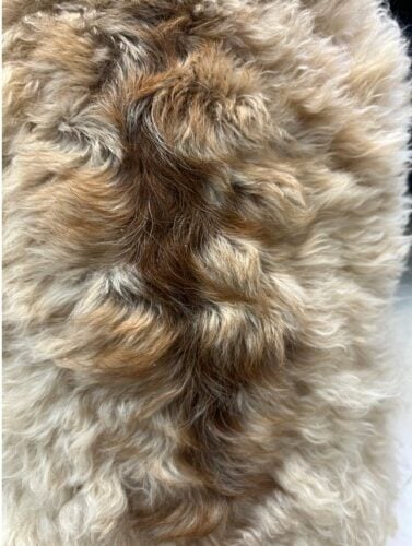
“Shaggy” presented for a 4-month history of progressive changes primarily to the hairs along the dorsal thoracic and caudothoracic trunk. No change was noted with antihistamines, lokivetmab (Cytopoint ®), nor melatonin.
.
Christie Yamazaki DVM, DACVD
Dermatology For Animals, Oakland, CA
February 2024
History:
“Shaggy” is a 1-year 8-month male neutered goldendoodle dog with a 4-month history of changes to the hair coat along the dorsal thorax. Mild pruritus was noted to the flanks, but it seemed to correlate with the end of the month, just before monthly parasiticides were due. There was a history of intermittent ear infections that resolved with a broad-spectrum antimicrobial otic treatment (Mometamax ™). “Shaggy” was obtained from a breeder at 10 weeks of age and had no other medical concerns or abnormalities noted. A complete blood count and biochemistry panel including thyroid hormone (T4) was performed with the referring veterinarian and all values were within normal limits. Melatonin 6 mg per os q12h appeared to be ineffective over the previous 3 months.
Exam:
Weight: 38.2 kg (84lb)
Temp: 101.4 F (rectal)
Pulse: 70 bpm
Resp: panting (eupneic)
Dermatologic physical examination: bright, alert, and responsive; Body Condition Score (BCS) 5/9. “Shaggy” was sweet and easily examined, though anxious. Hair coat is clean and overall curly, soft, blond-gold in color. There was mild tan cerumen in both ears, and both vertical and horizontal canals were clean and free from erythema. The tympanic membranes were challenging to fully visualize given the nervous temperament. There was a large serpiginous area of hairs that were straighter and darker gold than the hairs on the rest of the body. There was mild patchy peripheral hypotrichosis along the affected areas on the thoraco- and caudodorsum with mild patchy hyperpigmentation of the underlying skin. The paws had mild hypotrichosis and erythema of the palmar skin. There were no other dermatologic abnormalities. All lymph nodes were symmetrical on palpation with no obvious lymphadenomegaly.


Diagnostics:
Otic cytology was performed given the history of otitis externa and was unremarkable. Skin cytology was performed of the paws and revealed focal areas of abundant Malassezia yeasts. A strict elimination diet trial was recommended as the pododermatitis was considered likely secondary to either cutaneous adverse food reactions or atopic dermatitis.
Skin cytology and skin scrapes of the areas with affected hairs were unremarkable. A trichogram revealed notable follicular casts, which is keratosebaceous debris adhered to the hair shafts.
A percutaneous skin punch biopsy confirmed the diagnosis of follicular dysplasia. There was little to no inflammation detected in any of the tissue samples.
Assessment:
The clinical presentation of progressive non-inflammatory changes to the hair coat in this dog raised strong suspicion for follicular dysplasia. There is speculation of a genetic association with retriever-poodle crosses as the author and colleagues have seen a recent rise in doodles with nearly identical changes to the hairs in the same location. Further details to this condition are currently being investigated. Differential diagnoses include alopecia areata, canine flank alopecia, pyoderma secondary to parasite hypersensitivity, topical reactions secondary to contaminated shampoos or topical parasiticides, and endocrinopathy.
The unrelated historical recurrent episodes of otitis externa are likely secondary to a primary allergy and/or perpetuating or predisposing factors.
Plan:
Structural changes secondary to the dysplastic hair follicles predispose the hairs to structural damage such as fracture. The hairs that regrow often are weaker than the original hairs that were lost and are liable to recurrent trauma at areas of high friction or vigorous grooming. Changes often are mild at the initial onset but tend to progress, and alopecia can become permanent. These changes can predispose patients to secondary folliculitis, and so medicated topicals are often employed as prevention. In this case, a leave-on foam with antimicrobial properties (Douxo S3 Pyo Mousse®) was recommended for use at the affected areas on the back (as well as the paws).
Recommendations were made to further investigate for a concurrent primary allergic trigger.
Follow-up:
“Shaggy” had returned for a follow up visit 6 months later where hair regrowth was evident, though the color and textural changes persisted. Melatonin can be beneficial in some cases where estrogens are involved, so the family had continued supplementation without dramatic perceived benefit. Supplement was discontinued without appreciable change to the condition after 8 months. The allergic skin and ear diseases were considered to be unrelated to the changes in hair coat and continue to be managed by the author. Medicated bathing with a shampoo that contained antimicrobials as well as ceramides (Douxo S3 Pyo ®) was continued at weekly intervals.

Discussion:
Follicular dysplasia can be associated with specific coat colors given changes to the pigment (i.e. black hair follicular dysplasia, color dilution alopecia), and many have a genetic basis. Thus, no treatment prevents hair loss. Harsh grooming techniques or products (such as stripping the coat, benzoyl peroxide-based shampoos) can exacerbate the condition and/or result in secondary folliculitis and should be avoided.
Endocrine hair loss is the primary differential and more likely to become evident in later years of life, so it is important to rule this out in progressive or unresponsive cases.
References:
- Miller WH, Griffin CE, Campbell KL. Congenital and Hereditary Defects: Follicular Dysplasia. Muller and Kirk’s Small Animal Dermatology. 7th Philadelphia, PA: Saunders, 2013; 597-599.
Search terms
Follicular dysplasia dogs, follicular casting, clinical and histologic abnormalities, topical therapy, sebaceous glands, canine idiopathic sebaceous adenitis, conventional topical treatment, shaggy goldendoodle long hair, why is my goldendoodle losing hair, derma doodle, goldendoodle bald spot, conditions