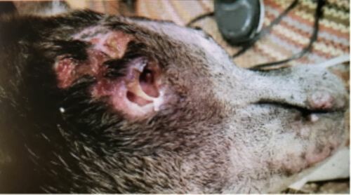
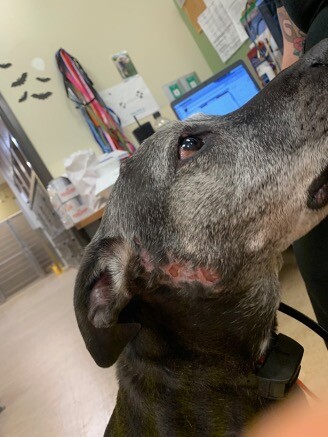
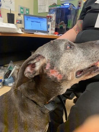
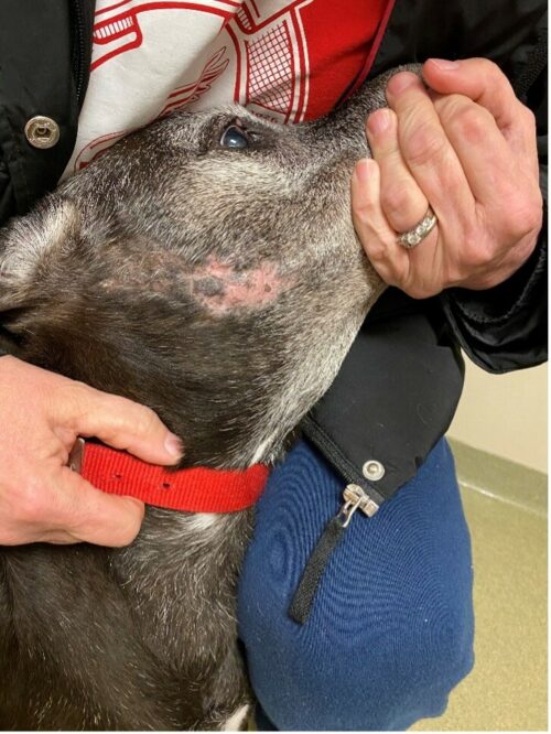
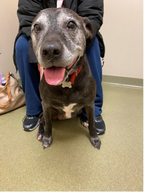
“Mia” is a 13y FS Mixed Breed Dog who presented for a refractory non-healing wound on the side of the face for 5 months prior to presentation.
Curtis Plowgian, DVM, DACVD
Animal Dermatology Clinic, Indianapolis, IN
January 2024
History:
The problem began as a raised red nodule/swelling on the side of the face, and at the time the primary veterinarian clipped and cleaned the wound and told the owner to treat it with topical Neosporin. Despite Neosporin and an ecollar, the lesion continued to worsen, became more widespread and swollen, and eventually draining tracts and fistulae developed. The owner saw the primary veterinarian a few months later, and the primary veterinarian started clindamycin, and after this was started, swelling improved, but the fistulae widened over time. In addition to the skin issues, Mia has a chronic history of IMHA, managed by an internist, that was currently well-controlled and stable on 8.6 mg/kg of mycophenolate BID. Mia was also on carprofen, tramadol and gabapentin for rear limb lameness (suspected cruciate injury). There were no other pets in the house, and the owner was unaffected. Mia had no other prior history of skin disease prior to this incident.
Exam:
On physical examination, the skin and coat were unremarkable except for a few soft, movable subcutaneous masses (suspected lipomas) over the flanks and ribs, and a large fistula on the right zygomatic region, surrounded by moist ulcerative dermatitis, and filled with purulent debris.
Diagnostics:
Cytology of the fistulae revealed TNTC white blood cells (primarily macrophages and neutrophils) with nuclear streaming, and TNTC intracellular and extracellular cocci with rare clusters of rods.
Bacterial culture and sensitivity grew a methicillin-resistant Staphylococcus pseudintermedius that was only sensitive to amikacin, chloramphenicol, and mupirocin, and a Pasteurella that was sensitive to several antibiotics including cephalosporins, fluoroquinolones, and sulfas, but resistant to chloramphenicol and mupirocin.
Assessment:
Pyogranulomatous fistulation of the zygomatic region with mixed bacterial infection, r/o secondary to infection/abscess vs sterile/immune-mediated process vs neoplasia vs tooth root abscess with external communication.
Plan:
While awaiting the culture results for the first 5 days, the owner was instructed to flush the wound copiously with cool running water for 5 minutes, prior to applying silver sulfadiazine cream. Dental radiographs were recommended with primary veterinarian to rule out tooth root involvement.
After culture results returned, it was discussed that given the mix of highly resistant staph with Pasteurella which was resistant to chloramphenicol, we had the options to pursue either amikacin or a combination of antibiotics. Given the cost and possible risk of adverse effects of amikacin, owner elected to try a combination of chloramphenicol (given at 34.1 mg/kg TID) and sulfamethoxazole-trimethoprim (given at 32.9 mg/kg BID), and to continue the previously prescribed hydrotherapy and silver sulfadiazine. A recheck was scheduled for 4 weeks.
Follow-up:
At 4 week recheck, Mia was markedly improved on the face, and the wound had closed up. Dental radiographs with the primary veterinarian had revealed no tooth root involvement.
In the last few days prior to the recheck, owner had noticed that Mia seemed to be getting weaker in her hind limbs. Repeat cytology showed rare white blood cells and no infectious organisms. The owner was instructed to stop the chloramphenicol, but continue the SMZ/TMP, hydrotherapy and SSD cream until recheck in 4 weeks. In 4 more weeks, the hind limb paresis had resolved, and the facial wound had further resolved:
Mia was able to discontinue antibiotics, and it was concluded that biopsy was not necessary to rule out immune-mediated disease or neoplasia. Mia was instructed to follow up with her primary veterinarian and internist about her stifle and IMHA management. She has not needed to return for further management of the skin.
Discussion:
This was an interesting case of how severe infection can present in the absence of an underlying cause such as neoplasia or autoimmune disease. It is unclear whether the initial infection was caused by a puncture (such as a bite wound, insect bite/sting, or splinter or other foreign body), or whether an infection or abscess developed secondary to immunosuppression from Mia’s IMHA, but given the lack of recurrence after antimicrobial therapy was discontinued, the author suspects that the former scenario is more likely than the latter.
In cases of deep infection (deep pyoderma, abscessation, granuloma), it is generally accepted that a minimum of 4-6 weeks of treatment with appropriate antimicrobials will be required, with treatment sometimes taking up to 12 weeks1. Antibiotics were selected by culture and sensitivity, which is appropriate in any cases that have failed to clear with antibiotics, and arguably appropriate in any cases where rods are found on infection (although the Staphylococcus turned out to be the much more resistant organism in this case). In the author’s opinion, it is also beneficial in the case of highly resistant infections to employ a multimodal approach to infection control, combining “inside out” treatment with oral antibiotics with an “outside in” topical therapy to enhance speed and efficacy of response.
This patient developed hind limb paresis on chloramphenicol, which is a documented side effect of this drug2, and the owner was warned to monitor for this when the medication was started. Hind limb weakness was shown to be the second most common side effect of this medication after GI upset, and was more common in large breed dogs than small breed dogs (mean weight 35.3kg)2. Given the fact that the period of treatment required to clear deep infections is often longer than 4 weeks, it was perhaps high risk to stop the chloramphenicol after 4 weeks, but given that the hind limb paresis is more likely to be reversible if the drug is continued quickly, and the fact that no bacteria was seen on follow up cytology, discontinuing of the drug was a judgment call that worked out in this case.
One other judgment call in this case that could be questioned in retrospect was the selection of sulfa antibiotics to treat the Pasturella, since this dog had a history of IMHA, and documented side effects of sulfa antibiotics include blood dyscrasias (including thrombocytopenia and hemolytic anemia)3. However, these adverse effects are rare and idiosyncratic, and the patient had been showing positive response to topical sulfas (silver sulfadiazine) without any adverse reactions, so the author felt that the potential benefits outweighed the risks, and again, it worked out in this case.
Pyoderma can present in many different ways, with different clinical signs, depth of infection, infectious organisms involved, and antimicrobial sensitivities. This case is a good example of the importance of culture and sensitivity, and multimodal therapy in the presence of highly resistant infections with multiple organisms.
References:
- Scott, D.W., Miller Jr, W.H., & Griffin, C.E. (2013). Muller and Kirk’s Small Animal Dermatology (7th). Elsevier, p 200.
- Short et al. Adverse events associated with chloramphenicol use in dogs: a retrospective study (2007–2013). Veterinary Record, Volume 175, Issue 21 p. 537-537.
- A. Trepanier. Idiosyncratic toxicity associated with potentiated sulfonamides in the dog. Journal of Veterinary Pharmacology and Therapeutics, Volume 27, Issue 3 p. 129-138
